sc-PDB
An Annotated Database of Druggable Binding Sites from the Protein DataBank
An Annotated Database of Druggable Binding Sites from the Protein DataBank
1.950 Å
X-ray
2013-03-22
| Name: | Putative FAD-dependent oxygenase EncM |
|---|---|
| ID: | Q9KHK2_9ACTN |
| AC: | Q9KHK2 |
| Organism: | Streptomyces maritimus |
| Reign: | Bacteria |
| TaxID: | 115828 |
| EC Number: | / |
| Chain Name: | Percentage of Residues within binding site |
|---|---|
| A | 100 % |
| B-Factor: | 11.295 |
|---|---|
| Number of residues: | 60 |
| Including | |
| Standard Amino Acids: | 54 |
| Non Standard Amino Acids: | 0 |
| Water Molecules: | 6 |
| Cofactors: | |
| Metals: | |
| Ligandability | Volume (Å3) |
|---|---|
| 1.188 | 1518.750 |
| % Hydrophobic | % Polar |
|---|---|
| 55.33 | 44.67 |
| According to VolSite | |
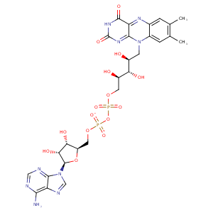
| HET Code: | FAD |
|---|---|
| Formula: | C27H31N9O15P2 |
| Molecular weight: | 783.534 g/mol |
| DrugBank ID: | DB03147 |
| Buried Surface Area: | 78.92 % |
| Polar Surface area: | 381.7 Å2 |
| Number of | |
|---|---|
| H-Bond Acceptors: | 22 |
| H-Bond Donors: | 7 |
| Rings: | 6 |
| Aromatic rings: | 3 |
| Anionic atoms: | 2 |
| Cationic atoms: | 0 |
| Rule of Five Violation: | 3 |
| Rotatable Bonds: | 13 |
| X | Y | Z |
|---|---|---|
| 12.0467 | 15.1753 | 36.3138 |


Represent the protein/ligand binding mode, centered on the ligand
Dashed lines represents hydrogen bonds and metal interactions
Green residue labels for amino acids with hydrophobic contacts (green lines) to the ligand
| Ligand | Protein | Interaction | |||
|---|---|---|---|---|---|
| Atom | Atom | Residue | Distance (Å) | Angle (°) | Type |
| C7M | CZ2 | TRP- 37 | 3.89 | 0 | Hydrophobic |
| C8M | CE3 | TRP- 37 | 3.22 | 0 | Hydrophobic |
| C2B | CB | VAL- 73 | 4.39 | 0 | Hydrophobic |
| O2A | N | GLY- 75 | 2.78 | 145.64 | H-Bond (Protein Donor) |
| O1P | N | GLY- 76 | 2.83 | 173.43 | H-Bond (Protein Donor) |
| O2A | N | GLY- 77 | 2.89 | 150.03 | H-Bond (Protein Donor) |
| C5' | CB | HIS- 78 | 3.51 | 0 | Hydrophobic |
| C8M | CB | HIS- 78 | 3.43 | 0 | Hydrophobic |
| O2P | N | HIS- 78 | 3.11 | 154.02 | H-Bond (Protein Donor) |
| O3P | N | HIS- 78 | 3.1 | 121.43 | H-Bond (Protein Donor) |
| O1A | N | SER- 79 | 2.86 | 150.61 | H-Bond (Protein Donor) |
| C5B | CB | SER- 79 | 4.05 | 0 | Hydrophobic |
| C1' | CE | MET- 80 | 4.27 | 0 | Hydrophobic |
| C9 | CE | MET- 80 | 3.53 | 0 | Hydrophobic |
| C3B | CB | HIS- 83 | 3.89 | 0 | Hydrophobic |
| O1A | OG | SER- 84 | 2.52 | 164.62 | H-Bond (Protein Donor) |
| N6A | O | GLY- 113 | 3.14 | 147.81 | H-Bond (Ligand Donor) |
| O4 | N | VAL- 135 | 3.11 | 156.14 | H-Bond (Protein Donor) |
| C6 | CG2 | VAL- 136 | 3.68 | 0 | Hydrophobic |
| C2' | CG2 | THR- 139 | 4.13 | 0 | Hydrophobic |
| C5' | CG2 | THR- 139 | 4.03 | 0 | Hydrophobic |
| C9A | CG2 | THR- 139 | 4.09 | 0 | Hydrophobic |
| O2P | N | GLY- 140 | 2.82 | 134.83 | H-Bond (Protein Donor) |
| O1P | N | GLY- 143 | 3.01 | 157 | H-Bond (Protein Donor) |
| C2' | CD1 | LEU- 144 | 4.37 | 0 | Hydrophobic |
| O2 | N | PHE- 150 | 2.96 | 162.83 | H-Bond (Protein Donor) |
| N3 | O | PHE- 150 | 2.8 | 149.81 | H-Bond (Ligand Donor) |
| N6A | O | VAL- 201 | 2.95 | 168.31 | H-Bond (Ligand Donor) |
| N1A | N | VAL- 201 | 2.92 | 154.07 | H-Bond (Protein Donor) |
| O2 | OH | TYR- 416 | 2.64 | 148.02 | H-Bond (Protein Donor) |
| C3' | CB | ASN- 418 | 3.75 | 0 | Hydrophobic |
| C1' | CE1 | PHE- 419 | 3.93 | 0 | Hydrophobic |
| O3B | O | LEU- 455 | 2.98 | 162.77 | H-Bond (Ligand Donor) |
| N5 | O | HOH- 794 | 2.94 | 170.19 | H-Bond (Protein Donor) |
| O2B | O | HOH- 827 | 2.69 | 156.48 | H-Bond (Ligand Donor) |
| O3' | O | HOH- 862 | 2.8 | 147.54 | H-Bond (Ligand Donor) |
