sc-PDB
An Annotated Database of Druggable Binding Sites from the Protein DataBank
An Annotated Database of Druggable Binding Sites from the Protein DataBank
1.500 Å
X-ray
2012-11-02
| Name: | Hygromycin-B 4-O-kinase |
|---|---|
| ID: | KHYB_ECOLX |
| AC: | P00557 |
| Organism: | Escherichia coli |
| Reign: | Bacteria |
| TaxID: | 562 |
| EC Number: | 2.7.1.163 |
| Chain Name: | Percentage of Residues within binding site |
|---|---|
| A | 100 % |
| B-Factor: | 20.118 |
|---|---|
| Number of residues: | 31 |
| Including | |
| Standard Amino Acids: | 27 |
| Non Standard Amino Acids: | 1 |
| Water Molecules: | 3 |
| Cofactors: | |
| Metals: | |
| Ligandability | Volume (Å3) |
|---|---|
| 1.198 | 448.875 |
| % Hydrophobic | % Polar |
|---|---|
| 57.89 | 42.11 |
| According to VolSite | |
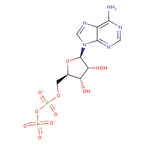
| HET Code: | ADP |
|---|---|
| Formula: | C10H12N5O10P2 |
| Molecular weight: | 424.177 g/mol |
| DrugBank ID: | - |
| Buried Surface Area: | 37.86 % |
| Polar Surface area: | 260.7 Å2 |
| Number of | |
|---|---|
| H-Bond Acceptors: | 14 |
| H-Bond Donors: | 3 |
| Rings: | 3 |
| Aromatic rings: | 2 |
| Anionic atoms: | 3 |
| Cationic atoms: | 0 |
| Rule of Five Violation: | 1 |
| Rotatable Bonds: | 6 |
| X | Y | Z |
|---|---|---|
| -51.1319 | 14.1246 | -24.8295 |
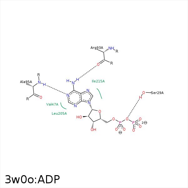


Represent the protein/ligand binding mode, centered on the ligand
Dashed lines represents hydrogen bonds and metal interactions
Green residue labels for amino acids with hydrophobic contacts (green lines) to the ligand
| Ligand | Protein | Interaction | |||
|---|---|---|---|---|---|
| Atom | Atom | Residue | Distance (Å) | Angle (°) | Type |
| C5' | CB | LEU- 28 | 3.87 | 0 | Hydrophobic |
| C4' | CD1 | LEU- 28 | 3.83 | 0 | Hydrophobic |
| O1B | OG | SER- 29 | 3.23 | 124.66 | H-Bond (Protein Donor) |
| O3A | OG | SER- 29 | 2.6 | 162.39 | H-Bond (Protein Donor) |
| C5' | CB | SER- 29 | 3.77 | 0 | Hydrophobic |
| C5' | CB | ALA- 36 | 4.05 | 0 | Hydrophobic |
| N6 | O | ARG- 93 | 2.96 | 138.37 | H-Bond (Ligand Donor) |
| N1 | N | ALA- 95 | 2.85 | 165.64 | H-Bond (Protein Donor) |
| C2' | CD1 | ILE- 215 | 4.49 | 0 | Hydrophobic |
