sc-PDB
An Annotated Database of Druggable Binding Sites from the Protein DataBank
An Annotated Database of Druggable Binding Sites from the Protein DataBank
1.390 Å
X-ray
2012-09-14
| Name: | DyP |
|---|---|
| ID: | Q8WZK8_9APHY |
| AC: | Q8WZK8 |
| Organism: | Bjerkandera adusta |
| Reign: | Eukaryota |
| TaxID: | 5331 |
| EC Number: | / |
| Chain Name: | Percentage of Residues within binding site |
|---|---|
| A | 100 % |
| B-Factor: | 10.254 |
|---|---|
| Number of residues: | 23 |
| Including | |
| Standard Amino Acids: | 21 |
| Non Standard Amino Acids: | 1 |
| Water Molecules: | 1 |
| Cofactors: | |
| Metals: | |
| Ligandability | Volume (Å3) |
|---|---|
| 1.112 | 816.750 |
| % Hydrophobic | % Polar |
|---|---|
| 47.11 | 52.89 |
| According to VolSite | |
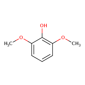
| HET Code: | 3DM |
|---|---|
| Formula: | C8H10O3 |
| Molecular weight: | 154.163 g/mol |
| DrugBank ID: | - |
| Buried Surface Area: | 63.12 % |
| Polar Surface area: | 38.69 Å2 |
| Number of | |
|---|---|
| H-Bond Acceptors: | 3 |
| H-Bond Donors: | 1 |
| Rings: | 1 |
| Aromatic rings: | 1 |
| Anionic atoms: | 0 |
| Cationic atoms: | 0 |
| Rule of Five Violation: | 0 |
| Rotatable Bonds: | 2 |
| X | Y | Z |
|---|---|---|
| -5.27573 | 7.96573 | 28.1835 |
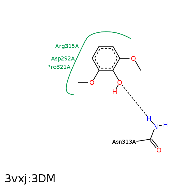


Represent the protein/ligand binding mode, centered on the ligand
Dashed lines represents hydrogen bonds and metal interactions
Green residue labels for amino acids with hydrophobic contacts (green lines) to the ligand
| Ligand | Protein | Interaction | |||
|---|---|---|---|---|---|
| Atom | Atom | Residue | Distance (Å) | Angle (°) | Type |
| C5 | CB | ASP- 292 | 4.03 | 0 | Hydrophobic |
| C8 | CB | ASP- 292 | 3.84 | 0 | Hydrophobic |
| O1 | ND2 | ASN- 313 | 3.14 | 161.99 | H-Bond (Protein Donor) |
| C8 | CD | ARG- 315 | 3.99 | 0 | Hydrophobic |
| C1 | CG | ARG- 315 | 3.68 | 0 | Hydrophobic |
| C4 | CB | ARG- 315 | 3.91 | 0 | Hydrophobic |
| C7 | CD1 | LEU- 318 | 3.77 | 0 | Hydrophobic |
| C7 | CB | VAL- 322 | 4.13 | 0 | Hydrophobic |
