sc-PDB
An Annotated Database of Druggable Binding Sites from the Protein DataBank
An Annotated Database of Druggable Binding Sites from the Protein DataBank
2.000 Å
X-ray
2011-09-21
| Name: | Hematopoietic prostaglandin D synthase |
|---|---|
| ID: | HPGDS_HUMAN |
| AC: | O60760 |
| Organism: | Homo sapiens |
| Reign: | Eukaryota |
| TaxID: | 9606 |
| EC Number: | / |
| Chain Name: | Percentage of Residues within binding site |
|---|---|
| B | 4 % |
| C | 96 % |
| B-Factor: | 20.203 |
|---|---|
| Number of residues: | 26 |
| Including | |
| Standard Amino Acids: | 24 |
| Non Standard Amino Acids: | 0 |
| Water Molecules: | 2 |
| Cofactors: | |
| Metals: | |
| Ligandability | Volume (Å3) |
|---|---|
| 1.229 | 394.875 |
| % Hydrophobic | % Polar |
|---|---|
| 60.68 | 39.32 |
| According to VolSite | |
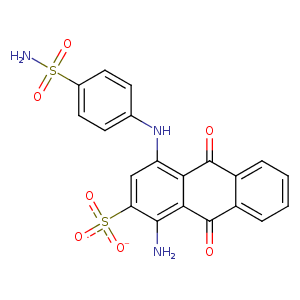
| HET Code: | M4M |
|---|---|
| Formula: | C20H14N3O7S2 |
| Molecular weight: | 472.471 g/mol |
| DrugBank ID: | - |
| Buried Surface Area: | 46.54 % |
| Polar Surface area: | 206.3 Å2 |
| Number of | |
|---|---|
| H-Bond Acceptors: | 9 |
| H-Bond Donors: | 3 |
| Rings: | 4 |
| Aromatic rings: | 3 |
| Anionic atoms: | 1 |
| Cationic atoms: | 0 |
| Rule of Five Violation: | 0 |
| Rotatable Bonds: | 4 |
| X | Y | Z |
|---|---|---|
| 0.832781 | -1.93316 | 97.4614 |
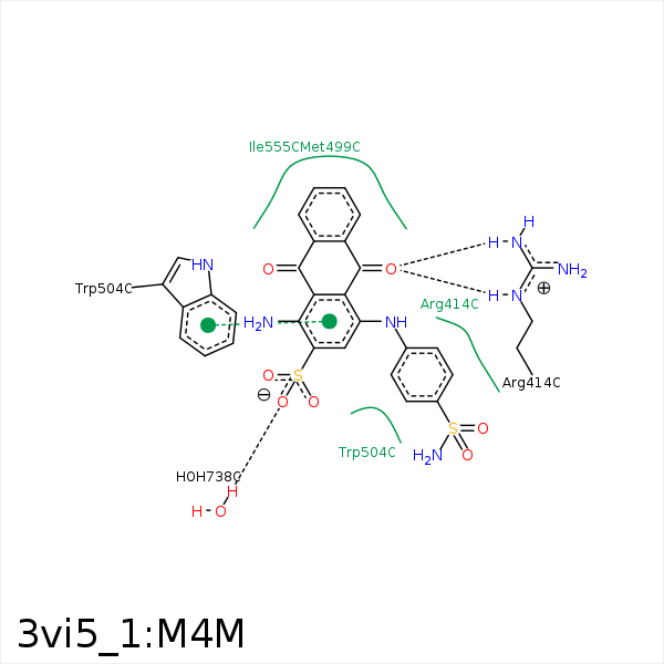


Represent the protein/ligand binding mode, centered on the ligand
Dashed lines represents hydrogen bonds and metal interactions
Green residue labels for amino acids with hydrophobic contacts (green lines) to the ligand
| Ligand | Protein | Interaction | |||
|---|---|---|---|---|---|
| Atom | Atom | Residue | Distance (Å) | Angle (°) | Type |
| O7 | NH2 | ARG- 414 | 2.79 | 126.78 | H-Bond (Protein Donor) |
| O7 | NE | ARG- 414 | 2.76 | 126.83 | H-Bond (Protein Donor) |
| C18 | CD | ARG- 414 | 3.9 | 0 | Hydrophobic |
| C1 | CG | ARG- 414 | 3.96 | 0 | Hydrophobic |
| C19 | CG | ARG- 414 | 3.91 | 0 | Hydrophobic |
| C3 | CG | MET- 499 | 3.66 | 0 | Hydrophobic |
| C2 | CB | MET- 499 | 3.56 | 0 | Hydrophobic |
| C4 | CE | MET- 499 | 3.82 | 0 | Hydrophobic |
| C14 | CB | TRP- 504 | 3.75 | 0 | Hydrophobic |
| C11 | CB | TRP- 504 | 3.41 | 0 | Hydrophobic |
| C4 | CG2 | ILE- 555 | 4.07 | 0 | Hydrophobic |
| C3 | SG | CYS- 556 | 3.7 | 0 | Hydrophobic |
| O3 | O | HOH- 738 | 2.91 | 147.26 | H-Bond (Protein Donor) |
