sc-PDB
An Annotated Database of Druggable Binding Sites from the Protein DataBank
An Annotated Database of Druggable Binding Sites from the Protein DataBank
1.200 Å
X-ray
2011-10-22
| Name: | Purine nucleoside phosphorylase DeoD-type |
|---|---|
| ID: | DEOD_BACCE |
| AC: | Q5EEL8 |
| Organism: | Bacillus cereus |
| Reign: | Bacteria |
| TaxID: | 1396 |
| EC Number: | / |
| Chain Name: | Percentage of Residues within binding site |
|---|---|
| A | 100 % |
| B-Factor: | 11.779 |
|---|---|
| Number of residues: | 28 |
| Including | |
| Standard Amino Acids: | 25 |
| Non Standard Amino Acids: | 0 |
| Water Molecules: | 3 |
| Cofactors: | |
| Metals: | |
| Ligandability | Volume (Å3) |
|---|---|
| 1.124 | 496.125 |
| % Hydrophobic | % Polar |
|---|---|
| 52.38 | 47.62 |
| According to VolSite | |
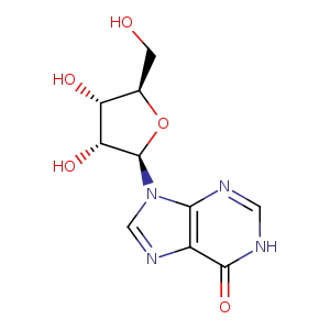
| HET Code: | NOS |
|---|---|
| Formula: | C10H12N4O5 |
| Molecular weight: | 268.226 g/mol |
| DrugBank ID: | DB04335 |
| Buried Surface Area: | 57.93 % |
| Polar Surface area: | 133.75 Å2 |
| Number of | |
|---|---|
| H-Bond Acceptors: | 8 |
| H-Bond Donors: | 4 |
| Rings: | 3 |
| Aromatic rings: | 2 |
| Anionic atoms: | 0 |
| Cationic atoms: | 0 |
| Rule of Five Violation: | 0 |
| Rotatable Bonds: | 2 |
| X | Y | Z |
|---|---|---|
| 43.9831 | 17.0516 | 42.1812 |
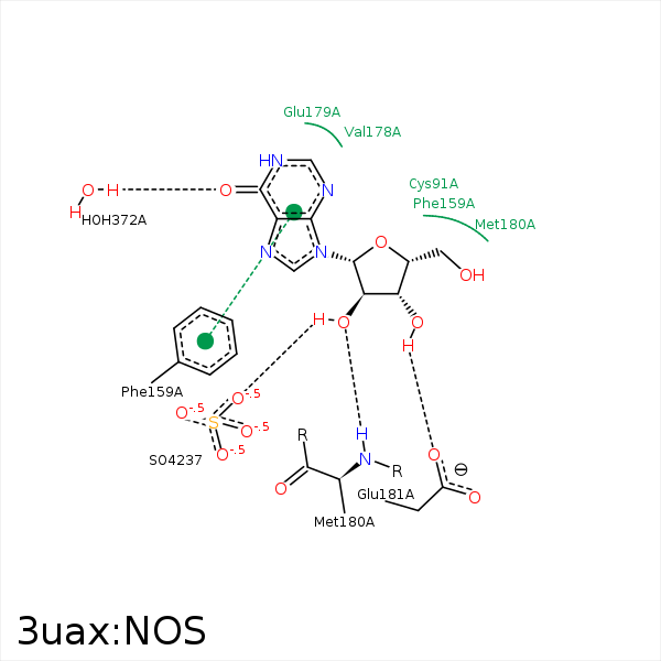


Represent the protein/ligand binding mode, centered on the ligand
Dashed lines represents hydrogen bonds and metal interactions
Green residue labels for amino acids with hydrophobic contacts (green lines) to the ligand
| Ligand | Protein | Interaction | |||
|---|---|---|---|---|---|
| Atom | Atom | Residue | Distance (Å) | Angle (°) | Type |
| C3' | CG | MET- 64 | 4.06 | 0 | Hydrophobic |
| C5' | SD | MET- 64 | 3.73 | 0 | Hydrophobic |
| C5' | CE2 | PHE- 159 | 3.72 | 0 | Hydrophobic |
| C2' | CB | GLU- 179 | 4.28 | 0 | Hydrophobic |
| C5' | SD | MET- 180 | 3.96 | 0 | Hydrophobic |
| C2' | CB | MET- 180 | 3.8 | 0 | Hydrophobic |
| O2' | N | MET- 180 | 2.85 | 128.57 | H-Bond (Protein Donor) |
| O3' | OE2 | GLU- 181 | 2.66 | 159.46 | H-Bond (Ligand Donor) |
| O6 | O | HOH- 372 | 2.6 | 179.96 | H-Bond (Protein Donor) |
