sc-PDB
An Annotated Database of Druggable Binding Sites from the Protein DataBank
An Annotated Database of Druggable Binding Sites from the Protein DataBank
2.000 Å
X-ray
2011-05-05
| Name: | UDP-N-acetylglucosamine 4-epimerase |
|---|---|
| ID: | GNE_PLESH |
| AC: | Q7BJX9 |
| Organism: | Plesiomonas shigelloides |
| Reign: | Bacteria |
| TaxID: | 703 |
| EC Number: | / |
| Chain Name: | Percentage of Residues within binding site |
|---|---|
| A | 100 % |
| B-Factor: | 35.359 |
|---|---|
| Number of residues: | 55 |
| Including | |
| Standard Amino Acids: | 52 |
| Non Standard Amino Acids: | 1 |
| Water Molecules: | 2 |
| Cofactors: | |
| Metals: | |
| Ligandability | Volume (Å3) |
|---|---|
| 0.992 | 1427.625 |
| % Hydrophobic | % Polar |
|---|---|
| 34.04 | 65.96 |
| According to VolSite | |
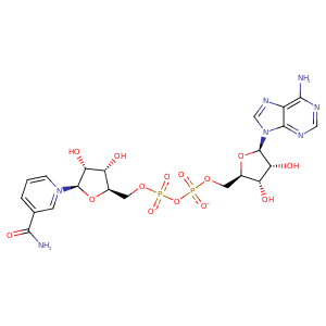
| HET Code: | NAD |
|---|---|
| Formula: | C21H26N7O14P2 |
| Molecular weight: | 662.417 g/mol |
| DrugBank ID: | - |
| Buried Surface Area: | 76.11 % |
| Polar Surface area: | 343.54 Å2 |
| Number of | |
|---|---|
| H-Bond Acceptors: | 18 |
| H-Bond Donors: | 6 |
| Rings: | 5 |
| Aromatic rings: | 3 |
| Anionic atoms: | 2 |
| Cationic atoms: | 1 |
| Rule of Five Violation: | 3 |
| Rotatable Bonds: | 11 |
| X | Y | Z |
|---|---|---|
| 8.93639 | 30.5645 | -11.0054 |
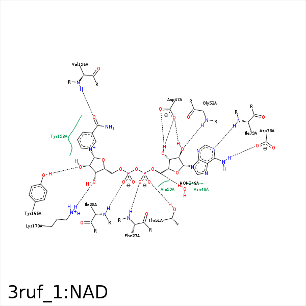


Represent the protein/ligand binding mode, centered on the ligand
Dashed lines represents hydrogen bonds and metal interactions
Green residue labels for amino acids with hydrophobic contacts (green lines) to the ligand
| Ligand | Protein | Interaction | |||
|---|---|---|---|---|---|
| Atom | Atom | Residue | Distance (Å) | Angle (°) | Type |
| O1A | N | PHE- 27 | 2.91 | 162.9 | H-Bond (Protein Donor) |
| O1N | N | ILE- 28 | 2.96 | 175.34 | H-Bond (Protein Donor) |
| C3N | CD1 | ILE- 28 | 4.49 | 0 | Hydrophobic |
| C5D | CD1 | ILE- 28 | 3.96 | 0 | Hydrophobic |
| O3B | OD2 | ASP- 47 | 2.79 | 135.02 | H-Bond (Ligand Donor) |
| O3B | OD1 | ASP- 47 | 3.23 | 158.37 | H-Bond (Ligand Donor) |
| O2B | OD1 | ASP- 47 | 2.56 | 157.87 | H-Bond (Ligand Donor) |
| N3A | N | ASN- 48 | 3.26 | 137.1 | H-Bond (Protein Donor) |
| O2B | OG | SER- 50 | 3.33 | 136.72 | H-Bond (Protein Donor) |
| O2A | OG1 | THR- 51 | 2.56 | 156.29 | H-Bond (Protein Donor) |
| O2B | N | THR- 51 | 3.43 | 121.41 | H-Bond (Protein Donor) |
| C2B | CB | THR- 51 | 4.44 | 0 | Hydrophobic |
| O2B | N | GLY- 52 | 2.8 | 138.05 | H-Bond (Protein Donor) |
| N6A | OD1 | ASP- 78 | 3.09 | 151.31 | H-Bond (Ligand Donor) |
| N1A | N | ILE- 79 | 3.1 | 169.19 | H-Bond (Protein Donor) |
| C5D | CB | GLN- 98 | 4.06 | 0 | Hydrophobic |
| C1B | CB | ALA- 99 | 4.36 | 0 | Hydrophobic |
| C3D | CB | ALA- 100 | 3.79 | 0 | Hydrophobic |
| C4D | CB | ALA- 140 | 3.67 | 0 | Hydrophobic |
| C5N | CB | SER- 142 | 4.12 | 0 | Hydrophobic |
| O2D | OH | TYR- 166 | 2.79 | 164.12 | H-Bond (Protein Donor) |
| O3D | NZ | LYS- 170 | 2.77 | 163.18 | H-Bond (Protein Donor) |
| O2D | NZ | LYS- 170 | 3.38 | 124.48 | H-Bond (Protein Donor) |
| C4D | CE1 | TYR- 193 | 4.45 | 0 | Hydrophobic |
| C5N | CB | TYR- 193 | 3.93 | 0 | Hydrophobic |
| C3N | CG2 | VAL- 196 | 4.47 | 0 | Hydrophobic |
| O7N | N | VAL- 196 | 3.05 | 153.52 | H-Bond (Protein Donor) |
| O5B | O | HOH- 348 | 3.13 | 171.07 | H-Bond (Protein Donor) |
