sc-PDB
An Annotated Database of Druggable Binding Sites from the Protein DataBank
An Annotated Database of Druggable Binding Sites from the Protein DataBank
2.000 Å
X-ray
2011-01-06
| Name: | N6 ADENINE SPECIFIC DNA METHYLASE (METHYLTRANSFERASE SUPERFAMILY) |
|---|---|
| ID: | Q8SRR4_ENCCU |
| AC: | Q8SRR4 |
| Organism: | Encephalitozoon cuniculi |
| Reign: | Eukaryota |
| TaxID: | 284813 |
| EC Number: | / |
| Chain Name: | Percentage of Residues within binding site |
|---|---|
| B | 100 % |
| B-Factor: | 20.738 |
|---|---|
| Number of residues: | 43 |
| Including | |
| Standard Amino Acids: | 40 |
| Non Standard Amino Acids: | 0 |
| Water Molecules: | 3 |
| Cofactors: | |
| Metals: | |
| Ligandability | Volume (Å3) |
|---|---|
| 0.813 | 550.125 |
| % Hydrophobic | % Polar |
|---|---|
| 51.53 | 48.47 |
| According to VolSite | |
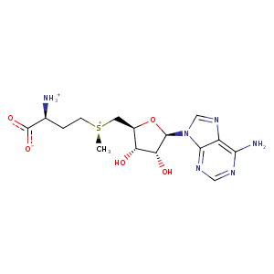
| HET Code: | SAM |
|---|---|
| Formula: | C15H23N6O5S |
| Molecular weight: | 399.445 g/mol |
| DrugBank ID: | DB00118 |
| Buried Surface Area: | 76.02 % |
| Polar Surface area: | 189.77 Å2 |
| Number of | |
|---|---|
| H-Bond Acceptors: | 9 |
| H-Bond Donors: | 4 |
| Rings: | 3 |
| Aromatic rings: | 2 |
| Anionic atoms: | 1 |
| Cationic atoms: | 2 |
| Rule of Five Violation: | 1 |
| Rotatable Bonds: | 7 |
| X | Y | Z |
|---|---|---|
| 36.4584 | 13.2884 | 13.3285 |
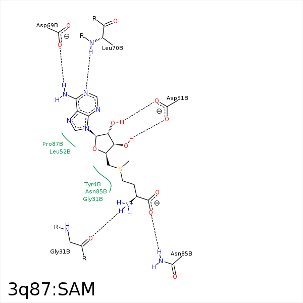


Represent the protein/ligand binding mode, centered on the ligand
Dashed lines represents hydrogen bonds and metal interactions
Green residue labels for amino acids with hydrophobic contacts (green lines) to the ligand
| Ligand | Protein | Interaction | |||
|---|---|---|---|---|---|
| Atom | Atom | Residue | Distance (Å) | Angle (°) | Type |
| SD | CG | TYR- 4 | 3.39 | 0 | Hydrophobic |
| CE | CE1 | TYR- 4 | 3.69 | 0 | Hydrophobic |
| C3' | CB | TYR- 4 | 4.34 | 0 | Hydrophobic |
| CG | CD2 | TYR- 4 | 3.66 | 0 | Hydrophobic |
| N | O | GLY- 31 | 2.7 | 162.99 | H-Bond (Ligand Donor) |
| CB | CB | SER- 33 | 3.58 | 0 | Hydrophobic |
| O3' | OD1 | ASP- 51 | 2.71 | 151.42 | H-Bond (Ligand Donor) |
| O2' | OD2 | ASP- 51 | 2.58 | 166.94 | H-Bond (Ligand Donor) |
| N3 | N | LEU- 52 | 3.45 | 140.83 | H-Bond (Protein Donor) |
| N6 | OD1 | ASP- 69 | 2.94 | 153.62 | H-Bond (Ligand Donor) |
| N1 | N | LEU- 70 | 2.89 | 172.11 | H-Bond (Protein Donor) |
| OXT | ND2 | ASN- 85 | 3.04 | 163.3 | H-Bond (Protein Donor) |
| CG | CB | ASN- 85 | 3.93 | 0 | Hydrophobic |
| C3' | CG2 | ILE- 96 | 4.06 | 0 | Hydrophobic |
| N | O | HOH- 171 | 3.44 | 176.11 | H-Bond (Ligand Donor) |
