sc-PDB
An Annotated Database of Druggable Binding Sites from the Protein DataBank
An Annotated Database of Druggable Binding Sites from the Protein DataBank
1.650 Å
X-ray
2010-12-09
| Name: | dTDP-3-amino-3,6-dideoxy-alpha-D-glucopyranose N,N-dimethyltransferase |
|---|---|
| ID: | TYLM1_STRFR |
| AC: | P95748 |
| Organism: | Streptomyces fradiae |
| Reign: | Bacteria |
| TaxID: | 1906 |
| EC Number: | 2.1.1.235 |
| Chain Name: | Percentage of Residues within binding site |
|---|---|
| D | 100 % |
| B-Factor: | 8.883 |
|---|---|
| Number of residues: | 37 |
| Including | |
| Standard Amino Acids: | 34 |
| Non Standard Amino Acids: | 1 |
| Water Molecules: | 2 |
| Cofactors: | |
| Metals: | |
| Ligandability | Volume (Å3) |
|---|---|
| 0.957 | 1069.875 |
| % Hydrophobic | % Polar |
|---|---|
| 47.00 | 53.00 |
| According to VolSite | |
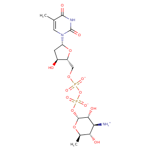
| HET Code: | T3Q |
|---|---|
| Formula: | C16H26N3O14P2 |
| Molecular weight: | 546.337 g/mol |
| DrugBank ID: | - |
| Buried Surface Area: | 67.44 % |
| Polar Surface area: | 283.77 Å2 |
| Number of | |
|---|---|
| H-Bond Acceptors: | 14 |
| H-Bond Donors: | 5 |
| Rings: | 3 |
| Aromatic rings: | 0 |
| Anionic atoms: | 2 |
| Cationic atoms: | 1 |
| Rule of Five Violation: | 2 |
| Rotatable Bonds: | 8 |
| X | Y | Z |
|---|---|---|
| -1.15009 | 2.11971 | 7.78049 |
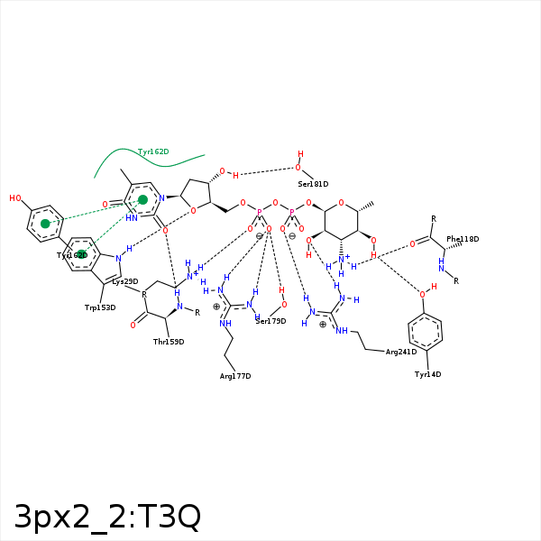


Represent the protein/ligand binding mode, centered on the ligand
Dashed lines represents hydrogen bonds and metal interactions
Green residue labels for amino acids with hydrophobic contacts (green lines) to the ligand
| Ligand | Protein | Interaction | |||
|---|---|---|---|---|---|
| Atom | Atom | Residue | Distance (Å) | Angle (°) | Type |
| O4Q | OH | TYR- 14 | 2.65 | 172.93 | H-Bond (Ligand Donor) |
| C4Q | CZ | TYR- 14 | 4.48 | 0 | Hydrophobic |
| O1A | NZ | LYS- 29 | 2.97 | 151.98 | H-Bond (Protein Donor) |
| O1A | NZ | LYS- 29 | 2.97 | 0 | Ionic (Protein Cationic) |
| O2B | NZ | LYS- 29 | 2.87 | 0 | Ionic (Protein Cationic) |
| N3Q | O | PHE- 118 | 3.17 | 152.43 | H-Bond (Ligand Donor) |
| C2Q | CZ | PHE- 118 | 4.3 | 0 | Hydrophobic |
| C1' | CH2 | TRP- 152 | 4.35 | 0 | Hydrophobic |
| C4' | CH2 | TRP- 152 | 3.79 | 0 | Hydrophobic |
| C2Q | CZ3 | TRP- 152 | 3.96 | 0 | Hydrophobic |
| C5M | CZ2 | TRP- 153 | 4.32 | 0 | Hydrophobic |
| C1' | CE2 | TRP- 153 | 3.85 | 0 | Hydrophobic |
| O4' | NE1 | TRP- 153 | 3 | 142.2 | H-Bond (Protein Donor) |
| C1' | CD1 | PHE- 158 | 4.41 | 0 | Hydrophobic |
| C2' | CE1 | PHE- 158 | 4.14 | 0 | Hydrophobic |
| O2 | N | THR- 159 | 3.02 | 168.99 | H-Bond (Protein Donor) |
| C5M | CZ | TYR- 162 | 3.92 | 0 | Hydrophobic |
| C5' | CE1 | TYR- 162 | 4.03 | 0 | Hydrophobic |
| C2' | CD1 | TYR- 162 | 3.58 | 0 | Hydrophobic |
| O2A | NH2 | ARG- 177 | 2.9 | 144.22 | H-Bond (Protein Donor) |
| O2A | NH1 | ARG- 177 | 2.83 | 148 | H-Bond (Protein Donor) |
| O2A | CZ | ARG- 177 | 3.31 | 0 | Ionic (Protein Cationic) |
| O3' | OG | SER- 181 | 2.7 | 156.44 | H-Bond (Ligand Donor) |
| C3' | CB | SER- 181 | 4.09 | 0 | Hydrophobic |
| C6Q | CG2 | ILE- 190 | 4.08 | 0 | Hydrophobic |
| C3' | CD1 | ILE- 190 | 4.06 | 0 | Hydrophobic |
| C6Q | CG2 | VAL- 192 | 4.37 | 0 | Hydrophobic |
| C4Q | CD1 | ILE- 212 | 4.45 | 0 | Hydrophobic |
| C2Q | CD1 | ILE- 212 | 4.46 | 0 | Hydrophobic |
| O1B | CZ | ARG- 241 | 3.87 | 0 | Ionic (Protein Cationic) |
| O1B | NH2 | ARG- 241 | 2.7 | 139 | H-Bond (Protein Donor) |
| O2Q | NH1 | ARG- 241 | 2.76 | 171.19 | H-Bond (Protein Donor) |
