sc-PDB
An Annotated Database of Druggable Binding Sites from the Protein DataBank
An Annotated Database of Druggable Binding Sites from the Protein DataBank
2.970 Å
X-ray
2010-12-07
| Name: | 1,2-phenylacetyl-CoA epoxidase, subunit A |
|---|---|
| ID: | PAAA_ECOLI |
| AC: | P76077 |
| Organism: | Escherichia coli |
| Reign: | Bacteria |
| TaxID: | 83333 |
| EC Number: | 1.14.13.149 |
| Chain Name: | Percentage of Residues within binding site |
|---|---|
| D | 100 % |
| B-Factor: | 43.129 |
|---|---|
| Number of residues: | 46 |
| Including | |
| Standard Amino Acids: | 46 |
| Non Standard Amino Acids: | 0 |
| Water Molecules: | 0 |
| Cofactors: | |
| Metals: | |
| Ligandability | Volume (Å3) |
|---|---|
| 0.770 | 867.375 |
| % Hydrophobic | % Polar |
|---|---|
| 37.35 | 62.65 |
| According to VolSite | |
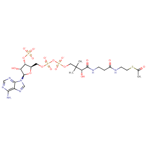
| HET Code: | ACO |
|---|---|
| Formula: | C23H34N7O17P3S |
| Molecular weight: | 805.539 g/mol |
| DrugBank ID: | - |
| Buried Surface Area: | 64.02 % |
| Polar Surface area: | 429.68 Å2 |
| Number of | |
|---|---|
| H-Bond Acceptors: | 22 |
| H-Bond Donors: | 5 |
| Rings: | 3 |
| Aromatic rings: | 2 |
| Anionic atoms: | 4 |
| Cationic atoms: | 0 |
| Rule of Five Violation: | 2 |
| Rotatable Bonds: | 20 |
| X | Y | Z |
|---|---|---|
| -14.4385 | -18.1941 | -36.2218 |
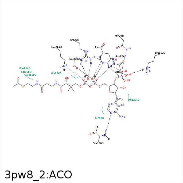


Represent the protein/ligand binding mode, centered on the ligand
Dashed lines represents hydrogen bonds and metal interactions
Green residue labels for amino acids with hydrophobic contacts (green lines) to the ligand
| Ligand | Protein | Interaction | |||
|---|---|---|---|---|---|
| Atom | Atom | Residue | Distance (Å) | Angle (°) | Type |
| O7A | CZ | ARG- 33 | 3.91 | 0 | Ionic (Protein Cationic) |
| O9A | CZ | ARG- 33 | 3.79 | 0 | Ionic (Protein Cationic) |
| O5A | CZ | ARG- 33 | 3.74 | 0 | Ionic (Protein Cationic) |
| O9A | NH1 | ARG- 33 | 2.7 | 153.15 | H-Bond (Protein Donor) |
| O5A | NH2 | ARG- 33 | 3.29 | 150.6 | H-Bond (Protein Donor) |
| O5A | NH1 | ARG- 33 | 3.25 | 152.69 | H-Bond (Protein Donor) |
| CAP | CG | GLN- 34 | 4.11 | 0 | Hydrophobic |
| O9A | NE2 | GLN- 37 | 2.8 | 151.92 | H-Bond (Protein Donor) |
| C6P | CB | HIS- 38 | 3.91 | 0 | Hydrophobic |
| S1P | CB | SER- 41 | 3.65 | 0 | Hydrophobic |
| C1B | CD | LYS- 103 | 3.89 | 0 | Hydrophobic |
| O8A | NZ | LYS- 103 | 2.74 | 166.1 | H-Bond (Protein Donor) |
| O8A | NZ | LYS- 103 | 2.74 | 0 | Ionic (Protein Cationic) |
| C2P | CB | SER- 105 | 4.1 | 0 | Hydrophobic |
| N1A | N | SER- 106 | 2.7 | 139.9 | H-Bond (Protein Donor) |
| N6A | OG | SER- 106 | 3.21 | 124.37 | H-Bond (Ligand Donor) |
| CH3 | CZ | PHE- 108 | 3.44 | 0 | Hydrophobic |
| CH3 | CG1 | VAL- 125 | 4.34 | 0 | Hydrophobic |
| CH3 | CB | ALA- 129 | 3.67 | 0 | Hydrophobic |
| C2P | CB | ALA- 129 | 3.86 | 0 | Hydrophobic |
| O9P | ND2 | ASN- 132 | 3.44 | 147.8 | H-Bond (Protein Donor) |
| N6A | O | MET- 193 | 3.43 | 126.95 | H-Bond (Ligand Donor) |
| N6A | O | MET- 194 | 3.37 | 132.89 | H-Bond (Ligand Donor) |
| C2P | CE | MET- 194 | 3.44 | 0 | Hydrophobic |
| CH3 | CE | MET- 194 | 4 | 0 | Hydrophobic |
| C5B | CG | PRO- 197 | 4.38 | 0 | Hydrophobic |
| O1A | OG | SER- 202 | 3.13 | 161.52 | H-Bond (Protein Donor) |
| C5B | CG | PRO- 203 | 4.03 | 0 | Hydrophobic |
| O7A | ND2 | ASN- 204 | 3.33 | 165.34 | H-Bond (Protein Donor) |
| O1A | N | ASN- 204 | 3.04 | 146.68 | H-Bond (Protein Donor) |
| O3A | ND2 | ASN- 204 | 2.98 | 166.35 | H-Bond (Protein Donor) |
| O2A | NZ | LYS- 214 | 3.72 | 0 | Ionic (Protein Cationic) |
| O4A | NZ | LYS- 214 | 2.66 | 0 | Ionic (Protein Cationic) |
| O4A | NZ | LYS- 214 | 2.66 | 160.13 | H-Bond (Protein Donor) |
| O2A | ND2 | ASN- 218 | 2.62 | 146.68 | H-Bond (Protein Donor) |
