sc-PDB
An Annotated Database of Druggable Binding Sites from the Protein DataBank
An Annotated Database of Druggable Binding Sites from the Protein DataBank
2.100 Å
X-ray
2010-12-07
| Name: | UDP-N-acetylglucosamine 4,6-dehydratase |
|---|---|
| ID: | Q5E8L1_VIBF1 |
| AC: | Q5E8L1 |
| Organism: | Vibrio fischeri |
| Reign: | Bacteria |
| TaxID: | 312309 |
| EC Number: | / |
| Chain Name: | Percentage of Residues within binding site |
|---|---|
| C | 94 % |
| D | 6 % |
| B-Factor: | 39.342 |
|---|---|
| Number of residues: | 50 |
| Including | |
| Standard Amino Acids: | 49 |
| Non Standard Amino Acids: | 0 |
| Water Molecules: | 1 |
| Cofactors: | |
| Metals: | |
| Ligandability | Volume (Å3) |
|---|---|
| 1.026 | 1134.000 |
| % Hydrophobic | % Polar |
|---|---|
| 40.48 | 59.52 |
| According to VolSite | |
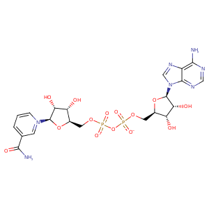
| HET Code: | NAD |
|---|---|
| Formula: | C21H26N7O14P2 |
| Molecular weight: | 662.417 g/mol |
| DrugBank ID: | - |
| Buried Surface Area: | 69.85 % |
| Polar Surface area: | 343.54 Å2 |
| Number of | |
|---|---|
| H-Bond Acceptors: | 18 |
| H-Bond Donors: | 6 |
| Rings: | 5 |
| Aromatic rings: | 3 |
| Anionic atoms: | 2 |
| Cationic atoms: | 1 |
| Rule of Five Violation: | 3 |
| Rotatable Bonds: | 11 |
| X | Y | Z |
|---|---|---|
| -0.699364 | 32.0148 | 65.1765 |


Represent the protein/ligand binding mode, centered on the ligand
Dashed lines represents hydrogen bonds and metal interactions
Green residue labels for amino acids with hydrophobic contacts (green lines) to the ligand
| Ligand | Protein | Interaction | |||
|---|---|---|---|---|---|
| Atom | Atom | Residue | Distance (Å) | Angle (°) | Type |
| O2A | OG | SER- 43 | 2.93 | 170.01 | H-Bond (Protein Donor) |
| O2A | N | SER- 43 | 2.93 | 164 | H-Bond (Protein Donor) |
| O2N | N | ILE- 44 | 2.9 | 176.91 | H-Bond (Protein Donor) |
| C5D | CG1 | ILE- 44 | 4.07 | 0 | Hydrophobic |
| O3B | OD2 | ASP- 64 | 2.74 | 158.32 | H-Bond (Ligand Donor) |
| O2B | OD1 | ASP- 64 | 2.6 | 157.91 | H-Bond (Ligand Donor) |
| C2B | CG2 | ILE- 65 | 4.41 | 0 | Hydrophobic |
| N3A | N | ILE- 65 | 3.34 | 147.35 | H-Bond (Protein Donor) |
| O1A | ND2 | ASN- 68 | 2.89 | 142.4 | H-Bond (Protein Donor) |
| N6A | OD1 | ASP- 94 | 2.81 | 149.81 | H-Bond (Ligand Donor) |
| N1A | N | ILE- 95 | 2.98 | 174.5 | H-Bond (Protein Donor) |
| C1B | CB | SER- 117 | 4.21 | 0 | Hydrophobic |
| O4B | N | ALA- 118 | 3.44 | 152.3 | H-Bond (Protein Donor) |
| C3D | CB | ALA- 118 | 3.54 | 0 | Hydrophobic |
| O3 | NZ | LYS- 120 | 3.45 | 129.73 | H-Bond (Protein Donor) |
| O1N | NZ | LYS- 120 | 2.9 | 156.86 | H-Bond (Protein Donor) |
| O1N | NZ | LYS- 120 | 2.9 | 0 | Ionic (Protein Cationic) |
| C2D | CB | LYS- 120 | 4.23 | 0 | Hydrophobic |
| C4D | CG2 | VAL- 160 | 4.07 | 0 | Hydrophobic |
| O3D | NZ | LYS- 176 | 2.7 | 153.53 | H-Bond (Protein Donor) |
| O2D | NZ | LYS- 176 | 2.79 | 120.44 | H-Bond (Protein Donor) |
| O7N | N | VAL- 200 | 2.86 | 153.55 | H-Bond (Protein Donor) |
| C3N | CG2 | VAL- 200 | 4.24 | 0 | Hydrophobic |
| N7N | OG | SER- 203 | 3 | 153.59 | H-Bond (Ligand Donor) |
| O5B | O | HOH- 400 | 3.15 | 180 | H-Bond (Protein Donor) |
