sc-PDB
An Annotated Database of Druggable Binding Sites from the Protein DataBank
An Annotated Database of Druggable Binding Sites from the Protein DataBank
2.070 Å
X-ray
2010-10-26
| Name: | Glyoxalate/3-oxopropanoate/4-oxobutanoate reductase |
|---|---|
| ID: | Q39R98_GEOMG |
| AC: | Q39R98 |
| Organism: | Geobacter metallireducens |
| Reign: | Bacteria |
| TaxID: | 269799 |
| EC Number: | / |
| Chain Name: | Percentage of Residues within binding site |
|---|---|
| B | 2 % |
| G | 98 % |
| B-Factor: | 41.357 |
|---|---|
| Number of residues: | 51 |
| Including | |
| Standard Amino Acids: | 48 |
| Non Standard Amino Acids: | 0 |
| Water Molecules: | 3 |
| Cofactors: | |
| Metals: | |
| Ligandability | Volume (Å3) |
|---|---|
| 0.907 | 1613.250 |
| % Hydrophobic | % Polar |
|---|---|
| 48.12 | 51.88 |
| According to VolSite | |
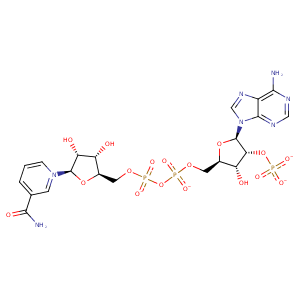
| HET Code: | NAP |
|---|---|
| Formula: | C21H25N7O17P3 |
| Molecular weight: | 740.381 g/mol |
| DrugBank ID: | DB03461 |
| Buried Surface Area: | 69.24 % |
| Polar Surface area: | 405.54 Å2 |
| Number of | |
|---|---|
| H-Bond Acceptors: | 21 |
| H-Bond Donors: | 5 |
| Rings: | 5 |
| Aromatic rings: | 3 |
| Anionic atoms: | 4 |
| Cationic atoms: | 1 |
| Rule of Five Violation: | 2 |
| Rotatable Bonds: | 13 |
| X | Y | Z |
|---|---|---|
| -7.36992 | 68.8551 | -35.1116 |


Represent the protein/ligand binding mode, centered on the ligand
Dashed lines represents hydrogen bonds and metal interactions
Green residue labels for amino acids with hydrophobic contacts (green lines) to the ligand
| Ligand | Protein | Interaction | |||
|---|---|---|---|---|---|
| Atom | Atom | Residue | Distance (Å) | Angle (°) | Type |
| O2A | N | ILE- 11 | 2.95 | 173.39 | H-Bond (Protein Donor) |
| O2N | N | MET- 12 | 2.91 | 170.22 | H-Bond (Protein Donor) |
| C5D | CB | MET- 12 | 4.49 | 0 | Hydrophobic |
| C3N | CG | MET- 12 | 3.57 | 0 | Hydrophobic |
| C4N | CE | MET- 12 | 3.47 | 0 | Hydrophobic |
| O3B | OD1 | ASN- 31 | 2.88 | 135.45 | H-Bond (Ligand Donor) |
| O3X | ND2 | ASN- 31 | 3.18 | 164 | H-Bond (Protein Donor) |
| O1X | CZ | ARG- 32 | 3.83 | 0 | Ionic (Protein Cationic) |
| O2X | CZ | ARG- 32 | 3.87 | 0 | Ionic (Protein Cationic) |
| O1X | OG | SER- 33 | 2.69 | 159.12 | H-Bond (Protein Donor) |
| O1X | N | SER- 33 | 3.07 | 123.14 | H-Bond (Protein Donor) |
| C5D | CG | MET- 64 | 4.07 | 0 | Hydrophobic |
| C4D | SD | MET- 64 | 3.96 | 0 | Hydrophobic |
| C1B | CD1 | LEU- 65 | 4.24 | 0 | Hydrophobic |
| O3D | O | LEU- 65 | 2.95 | 153.17 | H-Bond (Ligand Donor) |
| O4B | N | ALA- 66 | 3.1 | 128.11 | H-Bond (Protein Donor) |
| C5B | CB | ALA- 66 | 3.82 | 0 | Hydrophobic |
| N6A | OE2 | GLU- 73 | 3.14 | 168.28 | H-Bond (Ligand Donor) |
| O3D | N | THR- 96 | 3.18 | 149.96 | H-Bond (Protein Donor) |
| C2D | CB | THR- 96 | 4.36 | 0 | Hydrophobic |
| C5N | CG2 | VAL- 121 | 3.85 | 0 | Hydrophobic |
| O7N | OG | SER- 124 | 2.5 | 158.08 | H-Bond (Protein Donor) |
| N7N | O | ALA- 231 | 2.96 | 165.4 | H-Bond (Ligand Donor) |
| C2D | CE2 | PHE- 232 | 4.26 | 0 | Hydrophobic |
| O1A | NE2 | HIS- 236 | 3.03 | 172.12 | H-Bond (Protein Donor) |
| C2D | CB | HIS- 236 | 4.37 | 0 | Hydrophobic |
| O3D | NZ | LYS- 239 | 3.21 | 137.68 | H-Bond (Protein Donor) |
| O2N | O | HOH- 309 | 2.85 | 179.96 | H-Bond (Protein Donor) |
| O2D | O | HOH- 597 | 2.51 | 162.12 | H-Bond (Ligand Donor) |
