sc-PDB
An Annotated Database of Druggable Binding Sites from the Protein DataBank
An Annotated Database of Druggable Binding Sites from the Protein DataBank
1.200 Å
X-ray
2010-10-13
| Name: | Pentaerythritol tetranitrate reductase |
|---|---|
| ID: | P71278_ENTCL |
| AC: | P71278 |
| Organism: | Enterobacter cloacae |
| Reign: | Bacteria |
| TaxID: | 550 |
| EC Number: | / |
| Chain Name: | Percentage of Residues within binding site |
|---|---|
| A | 100 % |
| B-Factor: | 6.915 |
|---|---|
| Number of residues: | 20 |
| Including | |
| Standard Amino Acids: | 17 |
| Non Standard Amino Acids: | 2 |
| Water Molecules: | 1 |
| Cofactors: | FMN |
| Metals: | |
| Ligandability | Volume (Å3) |
|---|---|
| 0.323 | 300.375 |
| % Hydrophobic | % Polar |
|---|---|
| 33.71 | 66.29 |
| According to VolSite | |
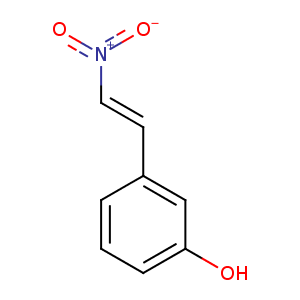
| HET Code: | P80 |
|---|---|
| Formula: | C8H7NO3 |
| Molecular weight: | 165.146 g/mol |
| DrugBank ID: | - |
| Buried Surface Area: | 62.11 % |
| Polar Surface area: | 66.05 Å2 |
| Number of | |
|---|---|
| H-Bond Acceptors: | 3 |
| H-Bond Donors: | 1 |
| Rings: | 1 |
| Aromatic rings: | 1 |
| Anionic atoms: | 1 |
| Cationic atoms: | 1 |
| Rule of Five Violation: | 0 |
| Rotatable Bonds: | 2 |
| X | Y | Z |
|---|---|---|
| 7.08808 | -11.311 | -24.0578 |
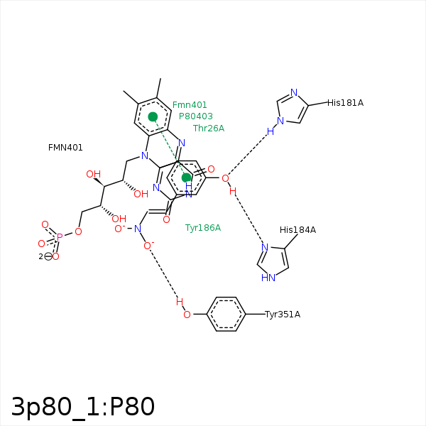


Represent the protein/ligand binding mode, centered on the ligand
Dashed lines represents hydrogen bonds and metal interactions
Green residue labels for amino acids with hydrophobic contacts (green lines) to the ligand
| Ligand | Protein | Interaction | |||
|---|---|---|---|---|---|
| Atom | Atom | Residue | Distance (Å) | Angle (°) | Type |
| C5' | CB | THR- 26 | 3.73 | 0 | Hydrophobic |
| O3' | NE2 | HIS- 181 | 2.74 | 154.21 | H-Bond (Protein Donor) |
| C3' | CB | HIS- 184 | 4.46 | 0 | Hydrophobic |
| O3' | ND1 | HIS- 184 | 2.7 | 165.04 | H-Bond (Ligand Donor) |
| O1 | OH | TYR- 351 | 2.63 | 139.41 | H-Bond (Protein Donor) |
| C2' | C1' | FMN- 401 | 4.02 | 0 | Hydrophobic |
