sc-PDB
An Annotated Database of Druggable Binding Sites from the Protein DataBank
An Annotated Database of Druggable Binding Sites from the Protein DataBank
1.540 Å
X-ray
2010-09-28
| Name: | Uridine phosphorylase 2 |
|---|---|
| ID: | UPP2_HUMAN |
| AC: | O95045 |
| Organism: | Homo sapiens |
| Reign: | Eukaryota |
| TaxID: | 9606 |
| EC Number: | 2.4.2.3 |
| Chain Name: | Percentage of Residues within binding site |
|---|---|
| A | 100 % |
| B-Factor: | 14.467 |
|---|---|
| Number of residues: | 24 |
| Including | |
| Standard Amino Acids: | 24 |
| Non Standard Amino Acids: | 0 |
| Water Molecules: | 0 |
| Cofactors: | |
| Metals: | |
| Ligandability | Volume (Å3) |
|---|---|
| 0.312 | 452.250 |
| % Hydrophobic | % Polar |
|---|---|
| 51.49 | 48.51 |
| According to VolSite | |
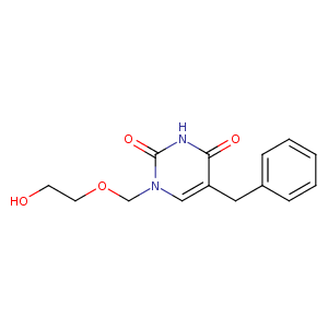
| HET Code: | BAU |
|---|---|
| Formula: | C14H16N2O4 |
| Molecular weight: | 276.288 g/mol |
| DrugBank ID: | DB07437 |
| Buried Surface Area: | 61.68 % |
| Polar Surface area: | 78.87 Å2 |
| Number of | |
|---|---|
| H-Bond Acceptors: | 4 |
| H-Bond Donors: | 2 |
| Rings: | 2 |
| Aromatic rings: | 1 |
| Anionic atoms: | 0 |
| Cationic atoms: | 0 |
| Rule of Five Violation: | 0 |
| Rotatable Bonds: | 6 |
| X | Y | Z |
|---|---|---|
| 13.9895 | -2.01795 | 57.8355 |
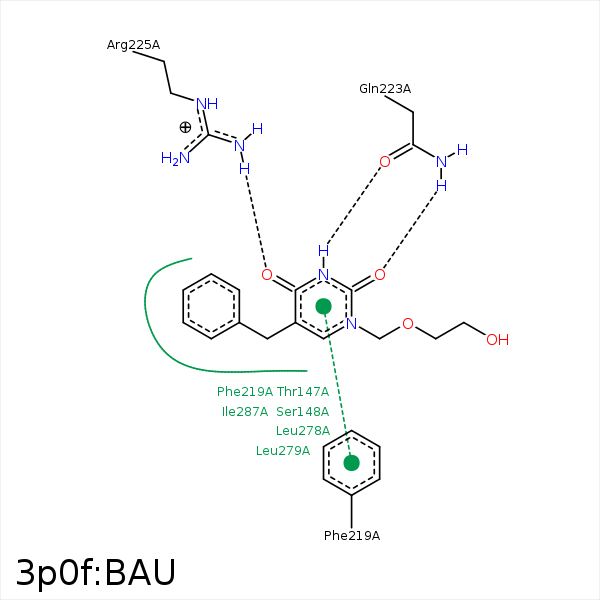


Represent the protein/ligand binding mode, centered on the ligand
Dashed lines represents hydrogen bonds and metal interactions
Green residue labels for amino acids with hydrophobic contacts (green lines) to the ligand
| Ligand | Protein | Interaction | |||
|---|---|---|---|---|---|
| Atom | Atom | Residue | Distance (Å) | Angle (°) | Type |
| CAJ | SD | MET- 116 | 3.42 | 0 | Hydrophobic |
| CAK | CE1 | PHE- 219 | 3.77 | 0 | Hydrophobic |
| OAB | NE2 | GLN- 223 | 3 | 170.98 | H-Bond (Protein Donor) |
| NAN | OE1 | GLN- 223 | 2.83 | 171.5 | H-Bond (Ligand Donor) |
| OAA | NH1 | ARG- 225 | 3.35 | 133.89 | H-Bond (Protein Donor) |
| OAA | NH2 | ARG- 225 | 2.84 | 163.9 | H-Bond (Protein Donor) |
| CAM | CB | GLU- 254 | 4.15 | 0 | Hydrophobic |
| CAK | SD | MET- 255 | 4.14 | 0 | Hydrophobic |
| CD2 | CD1 | LEU- 278 | 4.03 | 0 | Hydrophobic |
| CD1 | CD2 | LEU- 279 | 3.83 | 0 | Hydrophobic |
| CE1 | CG1 | ILE- 287 | 3.6 | 0 | Hydrophobic |
| CE2 | CD1 | ILE- 287 | 3.73 | 0 | Hydrophobic |
