sc-PDB
An Annotated Database of Druggable Binding Sites from the Protein DataBank
An Annotated Database of Druggable Binding Sites from the Protein DataBank
2.720 Å
X-ray
2010-09-01
| Name: | Dipeptidyl peptidase 4 |
|---|---|
| ID: | DPP4_HUMAN |
| AC: | P27487 |
| Organism: | Homo sapiens |
| Reign: | Eukaryota |
| TaxID: | 9606 |
| EC Number: | / |
| Chain Name: | Percentage of Residues within binding site |
|---|---|
| A | 100 % |
| B-Factor: | 27.128 |
|---|---|
| Number of residues: | 35 |
| Including | |
| Standard Amino Acids: | 33 |
| Non Standard Amino Acids: | 0 |
| Water Molecules: | 2 |
| Cofactors: | |
| Metals: | |
| Ligandability | Volume (Å3) |
|---|---|
| 0.000 | 374.625 |
| % Hydrophobic | % Polar |
|---|---|
| 30.63 | 69.37 |
| According to VolSite | |
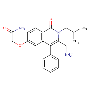
| HET Code: | LUI |
|---|---|
| Formula: | C22H26N3O3 |
| Molecular weight: | 380.460 g/mol |
| DrugBank ID: | - |
| Buried Surface Area: | 58.39 % |
| Polar Surface area: | 100.27 Å2 |
| Number of | |
|---|---|
| H-Bond Acceptors: | 3 |
| H-Bond Donors: | 2 |
| Rings: | 3 |
| Aromatic rings: | 2 |
| Anionic atoms: | 0 |
| Cationic atoms: | 1 |
| Rule of Five Violation: | 0 |
| Rotatable Bonds: | 7 |
| X | Y | Z |
|---|---|---|
| 17.8757 | -33.8962 | 14.7462 |
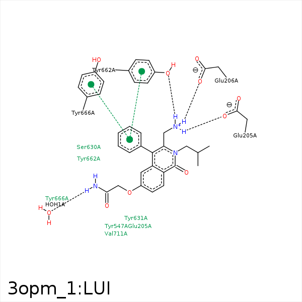


Represent the protein/ligand binding mode, centered on the ligand
Dashed lines represents hydrogen bonds and metal interactions
Green residue labels for amino acids with hydrophobic contacts (green lines) to the ligand
| Ligand | Protein | Interaction | |||
|---|---|---|---|---|---|
| Atom | Atom | Residue | Distance (Å) | Angle (°) | Type |
| N27 | O | HOH- 1 | 3.08 | 132.45 | H-Bond (Ligand Donor) |
| N18 | OE1 | GLU- 205 | 2.71 | 144.39 | H-Bond (Ligand Donor) |
| N18 | OE1 | GLU- 205 | 2.71 | 0 | Ionic (Ligand Cationic) |
| N18 | OE2 | GLU- 206 | 2.93 | 155.64 | H-Bond (Ligand Donor) |
| N18 | OE1 | GLU- 206 | 3.36 | 124.12 | H-Bond (Ligand Donor) |
| N18 | OE2 | GLU- 206 | 2.93 | 0 | Ionic (Ligand Cationic) |
| N18 | OE1 | GLU- 206 | 3.36 | 0 | Ionic (Ligand Cationic) |
| C22 | CE1 | PHE- 357 | 3.44 | 0 | Hydrophobic |
| C25 | CD1 | TYR- 547 | 3.69 | 0 | Hydrophobic |
| C4 | CB | SER- 630 | 4.09 | 0 | Hydrophobic |
| C13 | CB | SER- 630 | 3.74 | 0 | Hydrophobic |
| C15 | CB | TYR- 631 | 3.98 | 0 | Hydrophobic |
| C14 | CG2 | VAL- 656 | 4.37 | 0 | Hydrophobic |
| N18 | OH | TYR- 662 | 2.8 | 135.3 | H-Bond (Ligand Donor) |
| C13 | CZ | TYR- 662 | 3.45 | 0 | Hydrophobic |
| C15 | CZ | TYR- 666 | 3.5 | 0 | Hydrophobic |
| C13 | CG2 | VAL- 711 | 3.94 | 0 | Hydrophobic |
