sc-PDB
An Annotated Database of Druggable Binding Sites from the Protein DataBank
An Annotated Database of Druggable Binding Sites from the Protein DataBank
2.150 Å
X-ray
2010-08-17
| Name: | UDP-sugar pyrophosphorylase |
|---|---|
| ID: | D3G6S4_LEIMA |
| AC: | D3G6S4 |
| Organism: | Leishmania major |
| Reign: | Eukaryota |
| TaxID: | 5664 |
| EC Number: | / |
| Chain Name: | Percentage of Residues within binding site |
|---|---|
| A | 100 % |
| B-Factor: | 40.367 |
|---|---|
| Number of residues: | 35 |
| Including | |
| Standard Amino Acids: | 32 |
| Non Standard Amino Acids: | 0 |
| Water Molecules: | 3 |
| Cofactors: | |
| Metals: | |
| Ligandability | Volume (Å3) |
|---|---|
| 0.744 | 816.750 |
| % Hydrophobic | % Polar |
|---|---|
| 31.40 | 68.60 |
| According to VolSite | |
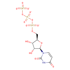
| HET Code: | UTP |
|---|---|
| Formula: | C9H11N2O15P3 |
| Molecular weight: | 480.109 g/mol |
| DrugBank ID: | DB04005 |
| Buried Surface Area: | 50.58 % |
| Polar Surface area: | 299.67 Å2 |
| Number of | |
|---|---|
| H-Bond Acceptors: | 15 |
| H-Bond Donors: | 3 |
| Rings: | 2 |
| Aromatic rings: | 0 |
| Anionic atoms: | 4 |
| Cationic atoms: | 0 |
| Rule of Five Violation: | 1 |
| Rotatable Bonds: | 8 |
| X | Y | Z |
|---|---|---|
| 9.72593 | 9.53455 | 29.0964 |
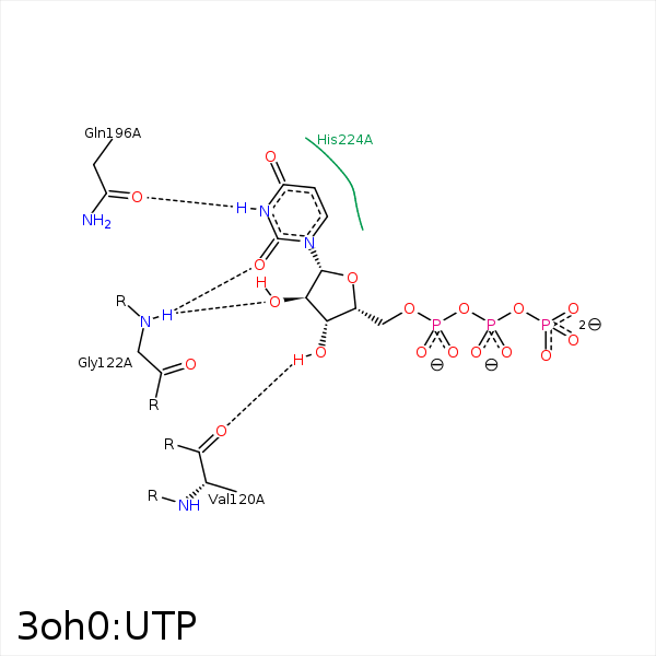


Represent the protein/ligand binding mode, centered on the ligand
Dashed lines represents hydrogen bonds and metal interactions
Green residue labels for amino acids with hydrophobic contacts (green lines) to the ligand
| Ligand | Protein | Interaction | |||
|---|---|---|---|---|---|
| Atom | Atom | Residue | Distance (Å) | Angle (°) | Type |
| C1' | CB | VAL- 120 | 4.43 | 0 | Hydrophobic |
| C3' | CG1 | VAL- 120 | 4 | 0 | Hydrophobic |
| O2' | N | GLY- 122 | 3.08 | 126.86 | H-Bond (Protein Donor) |
| O2 | N | GLY- 122 | 3.11 | 137.82 | H-Bond (Protein Donor) |
| O3G | NZ | LYS- 134 | 3.17 | 0 | Ionic (Protein Cationic) |
| N3 | OE1 | GLN- 196 | 2.79 | 165.14 | H-Bond (Ligand Donor) |
| O4 | NE2 | GLN- 196 | 3.33 | 123.58 | H-Bond (Protein Donor) |
| C1' | CB | HIS- 224 | 4.32 | 0 | Hydrophobic |
| C4' | CB | GLN- 270 | 4.44 | 0 | Hydrophobic |
| C3' | CB | ASP- 271 | 4.17 | 0 | Hydrophobic |
| O3' | N | ASP- 271 | 3.46 | 121.61 | H-Bond (Protein Donor) |
