sc-PDB
An Annotated Database of Druggable Binding Sites from the Protein DataBank
An Annotated Database of Druggable Binding Sites from the Protein DataBank
1.500 Å
X-ray
2010-08-04
| Name: | UDP-N-acetyl-2-amino-2-deoxy-D-glucuronate oxidase |
|---|---|
| ID: | WBPB_PSEAE |
| AC: | G3XD23 |
| Organism: | Pseudomonas aeruginosa |
| Reign: | Bacteria |
| TaxID: | 208964 |
| EC Number: | / |
| Chain Name: | Percentage of Residues within binding site |
|---|---|
| A | 9 % |
| C | 86 % |
| D | 5 % |
| B-Factor: | 15.562 |
|---|---|
| Number of residues: | 47 |
| Including | |
| Standard Amino Acids: | 43 |
| Non Standard Amino Acids: | 0 |
| Water Molecules: | 4 |
| Cofactors: | |
| Metals: | |
| Ligandability | Volume (Å3) |
|---|---|
| 0.651 | 1829.250 |
| % Hydrophobic | % Polar |
|---|---|
| 43.54 | 56.46 |
| According to VolSite | |
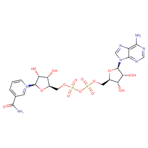
| HET Code: | NAD |
|---|---|
| Formula: | C21H26N7O14P2 |
| Molecular weight: | 662.417 g/mol |
| DrugBank ID: | - |
| Buried Surface Area: | 71.37 % |
| Polar Surface area: | 343.54 Å2 |
| Number of | |
|---|---|
| H-Bond Acceptors: | 18 |
| H-Bond Donors: | 6 |
| Rings: | 5 |
| Aromatic rings: | 3 |
| Anionic atoms: | 2 |
| Cationic atoms: | 1 |
| Rule of Five Violation: | 3 |
| Rotatable Bonds: | 11 |
| X | Y | Z |
|---|---|---|
| 65.2454 | 23.709 | 30.5727 |
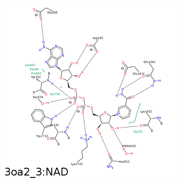


Represent the protein/ligand binding mode, centered on the ligand
Dashed lines represents hydrogen bonds and metal interactions
Green residue labels for amino acids with hydrophobic contacts (green lines) to the ligand
| Ligand | Protein | Interaction | |||
|---|---|---|---|---|---|
| Atom | Atom | Residue | Distance (Å) | Angle (°) | Type |
| O2A | N | TYR- 12 | 2.83 | 166.77 | H-Bond (Protein Donor) |
| O2N | N | ILE- 13 | 2.95 | 175.32 | H-Bond (Protein Donor) |
| C5D | CG2 | ILE- 13 | 3.83 | 0 | Hydrophobic |
| C4N | CD1 | ILE- 13 | 3.59 | 0 | Hydrophobic |
| O3B | OD1 | ASP- 33 | 3.43 | 126.98 | H-Bond (Ligand Donor) |
| O3B | OD2 | ASP- 33 | 2.68 | 179.07 | H-Bond (Ligand Donor) |
| O2B | OD1 | ASP- 33 | 2.7 | 155.1 | H-Bond (Ligand Donor) |
| O2A | OG | SER- 37 | 2.66 | 153.23 | H-Bond (Protein Donor) |
| N6A | OE1 | GLU- 55 | 3.02 | 162.84 | H-Bond (Ligand Donor) |
| O3D | OD1 | ASN- 81 | 2.75 | 161.75 | H-Bond (Ligand Donor) |
| C4D | CB | GLU- 101 | 4.13 | 0 | Hydrophobic |
| N7N | OE1 | GLU- 101 | 2.78 | 161.84 | H-Bond (Ligand Donor) |
| O2D | O | LYS- 102 | 2.79 | 159.27 | H-Bond (Ligand Donor) |
| O7N | NE2 | GLN- 130 | 3.07 | 159.58 | H-Bond (Protein Donor) |
| N7N | OE1 | GLN- 130 | 3.24 | 167.03 | H-Bond (Ligand Donor) |
| O1A | NE1 | TRP- 171 | 2.79 | 161.81 | H-Bond (Protein Donor) |
| C5D | CZ2 | TRP- 171 | 4.39 | 0 | Hydrophobic |
| C3D | CH2 | TRP- 171 | 3.8 | 0 | Hydrophobic |
| O1N | NZ | LYS- 172 | 2.81 | 172.05 | H-Bond (Protein Donor) |
| O1N | NZ | LYS- 172 | 2.81 | 0 | Ionic (Protein Cationic) |
| O5B | O | HOH- 352 | 3.39 | 150.37 | H-Bond (Protein Donor) |
| O2D | O | HOH- 416 | 2.7 | 151.64 | H-Bond (Protein Donor) |
| O2D | O | HOH- 423 | 3.38 | 120.15 | H-Bond (Protein Donor) |
