sc-PDB
An Annotated Database of Druggable Binding Sites from the Protein DataBank
An Annotated Database of Druggable Binding Sites from the Protein DataBank
2.530 Å
X-ray
2010-07-25
| Name: | Glycosyl transferase, family 2 |
|---|---|
| ID: | Q1ATN7_RUBXD |
| AC: | Q1ATN7 |
| Organism: | Rubrobacter xylanophilus |
| Reign: | Bacteria |
| TaxID: | 266117 |
| EC Number: | / |
| Chain Name: | Percentage of Residues within binding site |
|---|---|
| A | 100 % |
| B-Factor: | 39.532 |
|---|---|
| Number of residues: | 46 |
| Including | |
| Standard Amino Acids: | 43 |
| Non Standard Amino Acids: | 1 |
| Water Molecules: | 2 |
| Cofactors: | |
| Metals: | MG |
| Ligandability | Volume (Å3) |
|---|---|
| 0.698 | 840.375 |
| % Hydrophobic | % Polar |
|---|---|
| 36.95 | 63.05 |
| According to VolSite | |
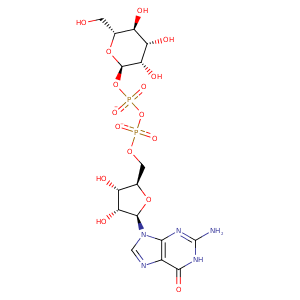
| HET Code: | GDD |
|---|---|
| Formula: | C16H23N5O16P2 |
| Molecular weight: | 603.325 g/mol |
| DrugBank ID: | - |
| Buried Surface Area: | 69.4 % |
| Polar Surface area: | 352.71 Å2 |
| Number of | |
|---|---|
| H-Bond Acceptors: | 19 |
| H-Bond Donors: | 8 |
| Rings: | 4 |
| Aromatic rings: | 1 |
| Anionic atoms: | 2 |
| Cationic atoms: | 0 |
| Rule of Five Violation: | 3 |
| Rotatable Bonds: | 9 |
| X | Y | Z |
|---|---|---|
| 43.8234 | -23.5594 | -5.58987 |
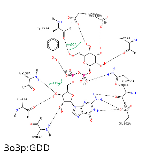


Represent the protein/ligand binding mode, centered on the ligand
Dashed lines represents hydrogen bonds and metal interactions
Green residue labels for amino acids with hydrophobic contacts (green lines) to the ligand
| Ligand | Protein | Interaction | |||
|---|---|---|---|---|---|
| Atom | Atom | Residue | Distance (Å) | Angle (°) | Type |
| C1' | CB | PRO- 49 | 4.36 | 0 | Hydrophobic |
| O3' | O | PRO- 49 | 3.05 | 169.38 | H-Bond (Ligand Donor) |
| C1' | CB | ARG- 51 | 4.45 | 0 | Hydrophobic |
| O2' | N | ARG- 51 | 2.97 | 140.5 | H-Bond (Protein Donor) |
| N2 | O | VAL- 80 | 3.01 | 138.54 | H-Bond (Ligand Donor) |
| N2 | OE1 | GLU- 102 | 2.95 | 134.41 | H-Bond (Ligand Donor) |
| N1 | OE1 | GLU- 102 | 2.73 | 150.58 | H-Bond (Ligand Donor) |
| C1' | CG | LYS- 115 | 4.42 | 0 | Hydrophobic |
| C31 | CD | LYS- 115 | 4.4 | 0 | Hydrophobic |
| C5' | CD | LYS- 115 | 4.19 | 0 | Hydrophobic |
| O31 | NZ | LYS- 115 | 3.13 | 122.08 | H-Bond (Protein Donor) |
| C4' | CB | ASP- 135 | 4.4 | 0 | Hydrophobic |
| O31 | OD2 | ASP- 135 | 3.37 | 173.3 | H-Bond (Ligand Donor) |
| O3' | N | ALA- 136 | 2.98 | 145.57 | H-Bond (Protein Donor) |
| C3' | CB | ASP- 137 | 4.23 | 0 | Hydrophobic |
| O21 | O | LEU- 207 | 3.01 | 154.8 | H-Bond (Ligand Donor) |
| C61 | CB | LEU- 207 | 3.74 | 0 | Hydrophobic |
| O1A | OH | TYR- 227 | 2.67 | 137.05 | H-Bond (Protein Donor) |
| C51 | CD1 | TYR- 227 | 4.48 | 0 | Hydrophobic |
| O6A | O | TYR- 227 | 2.93 | 171.93 | H-Bond (Ligand Donor) |
| O41 | OE2 | GLU- 230 | 3.03 | 143.23 | H-Bond (Ligand Donor) |
| C41 | CG | GLU- 230 | 3.59 | 0 | Hydrophobic |
| O2B | ND2 | ASN- 256 | 3.23 | 137.21 | H-Bond (Protein Donor) |
| O3B | NE2 | GLN- 259 | 2.65 | 153.9 | H-Bond (Protein Donor) |
| C51 | CE | MET- 267 | 3.83 | 0 | Hydrophobic |
| O2A | MG | MG- 339 | 2.69 | 0 | Metal Acceptor |
| O2B | MG | MG- 339 | 2.33 | 0 | Metal Acceptor |
