sc-PDB
An Annotated Database of Druggable Binding Sites from the Protein DataBank
An Annotated Database of Druggable Binding Sites from the Protein DataBank
2.010 Å
X-ray
2010-07-12
| Name: | Ferulic acid decarboxylase |
|---|---|
| ID: | C6F3U5_9ENTR |
| AC: | C6F3U5 |
| Organism: | Enterobacter sp. Px6-4 |
| Reign: | Bacteria |
| TaxID: | 418698 |
| EC Number: | / |
| Chain Name: | Percentage of Residues within binding site |
|---|---|
| A | 100 % |
| B-Factor: | 25.897 |
|---|---|
| Number of residues: | 18 |
| Including | |
| Standard Amino Acids: | 16 |
| Non Standard Amino Acids: | 0 |
| Water Molecules: | 2 |
| Cofactors: | |
| Metals: | |
| Ligandability | Volume (Å3) |
|---|---|
| 1.282 | 459.000 |
| % Hydrophobic | % Polar |
|---|---|
| 61.76 | 38.24 |
| According to VolSite | |
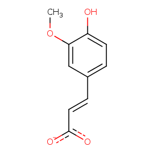
| HET Code: | FER |
|---|---|
| Formula: | C10H9O4 |
| Molecular weight: | 193.176 g/mol |
| DrugBank ID: | DB07767 |
| Buried Surface Area: | 51.67 % |
| Polar Surface area: | 69.59 Å2 |
| Number of | |
|---|---|
| H-Bond Acceptors: | 4 |
| H-Bond Donors: | 1 |
| Rings: | 1 |
| Aromatic rings: | 1 |
| Anionic atoms: | 1 |
| Cationic atoms: | 0 |
| Rule of Five Violation: | 0 |
| Rotatable Bonds: | 3 |
| X | Y | Z |
|---|---|---|
| 8.78736 | 10.5139 | 12.5319 |
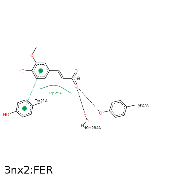


Represent the protein/ligand binding mode, centered on the ligand
Dashed lines represents hydrogen bonds and metal interactions
Green residue labels for amino acids with hydrophobic contacts (green lines) to the ligand
| Ligand | Protein | Interaction | |||
|---|---|---|---|---|---|
| Atom | Atom | Residue | Distance (Å) | Angle (°) | Type |
| C5 | CG | TYR- 21 | 3.36 | 0 | Hydrophobic |
| C5 | CB | TYR- 21 | 4.23 | 0 | Hydrophobic |
| O2 | OH | TYR- 27 | 2.94 | 159.56 | H-Bond (Protein Donor) |
| C2 | CG2 | ILE- 132 | 4.17 | 0 | Hydrophobic |
| O2 | O | HOH- 284 | 2.98 | 150.3 | H-Bond (Protein Donor) |
