sc-PDB
An Annotated Database of Druggable Binding Sites from the Protein DataBank
An Annotated Database of Druggable Binding Sites from the Protein DataBank
1.700 Å
X-ray
2010-06-14
| Name: | Ribosyldihydronicotinamide dehydrogenase [quinone] |
|---|---|
| ID: | NQO2_HUMAN |
| AC: | P16083 |
| Organism: | Homo sapiens |
| Reign: | Eukaryota |
| TaxID: | 9606 |
| EC Number: | / |
| Chain Name: | Percentage of Residues within binding site |
|---|---|
| A | 40 % |
| B | 60 % |
| B-Factor: | 27.981 |
|---|---|
| Number of residues: | 26 |
| Including | |
| Standard Amino Acids: | 24 |
| Non Standard Amino Acids: | 1 |
| Water Molecules: | 1 |
| Cofactors: | FAD |
| Metals: | |
| Ligandability | Volume (Å3) |
|---|---|
| 0.774 | 718.875 |
| % Hydrophobic | % Polar |
|---|---|
| 46.48 | 53.52 |
| According to VolSite | |
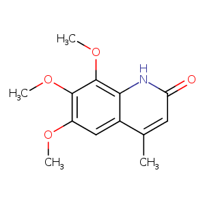
| HET Code: | HGZ |
|---|---|
| Formula: | C13H15NO4 |
| Molecular weight: | 249.262 g/mol |
| DrugBank ID: | - |
| Buried Surface Area: | 63.58 % |
| Polar Surface area: | 56.79 Å2 |
| Number of | |
|---|---|
| H-Bond Acceptors: | 4 |
| H-Bond Donors: | 1 |
| Rings: | 2 |
| Aromatic rings: | 1 |
| Anionic atoms: | 0 |
| Cationic atoms: | 0 |
| Rule of Five Violation: | 0 |
| Rotatable Bonds: | 3 |
| X | Y | Z |
|---|---|---|
| 23.3571 | -16.713 | -14.6258 |
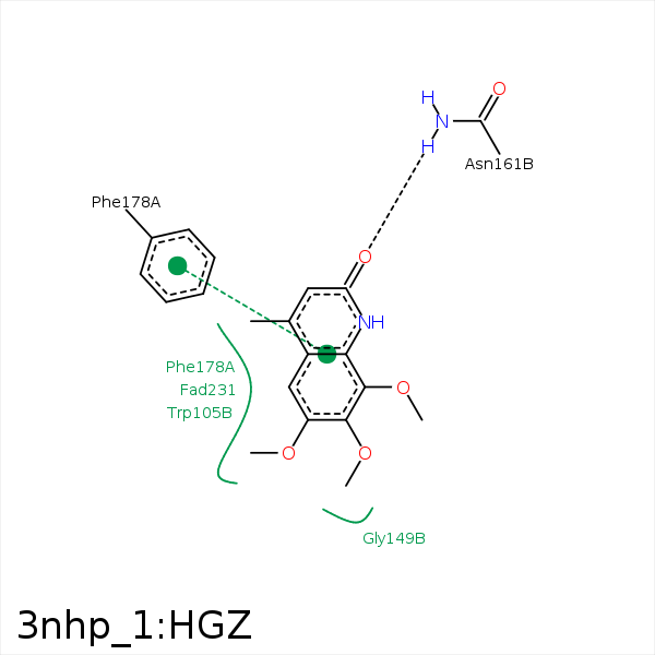


Represent the protein/ligand binding mode, centered on the ligand
Dashed lines represents hydrogen bonds and metal interactions
Green residue labels for amino acids with hydrophobic contacts (green lines) to the ligand
| Ligand | Protein | Interaction | |||
|---|---|---|---|---|---|
| Atom | Atom | Residue | Distance (Å) | Angle (°) | Type |
| C12 | CZ3 | TRP- 105 | 3.84 | 0 | Hydrophobic |
| C18 | CZ2 | TRP- 105 | 3.38 | 0 | Hydrophobic |
| C12 | CE1 | PHE- 106 | 3.8 | 0 | Hydrophobic |
| C18 | CZ | PHE- 126 | 3.28 | 0 | Hydrophobic |
| O11 | ND2 | ASN- 161 | 2.65 | 157.87 | H-Bond (Protein Donor) |
| C12 | CD1 | PHE- 178 | 3.49 | 0 | Hydrophobic |
| C16 | C9 | FAD- 231 | 4.07 | 0 | Hydrophobic |
| C18 | C6 | FAD- 231 | 3.39 | 0 | Hydrophobic |
| C5 | C1' | FAD- 231 | 3.7 | 0 | Hydrophobic |
