sc-PDB
An Annotated Database of Druggable Binding Sites from the Protein DataBank
An Annotated Database of Druggable Binding Sites from the Protein DataBank
1.600 Å
X-ray
2010-05-14
| Name: | Adenylate cyclase 2 |
|---|---|
| ID: | Q7CH76_YERPE |
| AC: | Q7CH76 |
| Organism: | Yersinia pestis |
| Reign: | Bacteria |
| TaxID: | 632 |
| EC Number: | / |
| Chain Name: | Percentage of Residues within binding site |
|---|---|
| B | 100 % |
| B-Factor: | 26.049 |
|---|---|
| Number of residues: | 29 |
| Including | |
| Standard Amino Acids: | 28 |
| Non Standard Amino Acids: | 1 |
| Water Molecules: | 0 |
| Cofactors: | |
| Metals: | MN |
| Ligandability | Volume (Å3) |
|---|---|
| 0.893 | 1161.000 |
| % Hydrophobic | % Polar |
|---|---|
| 38.37 | 61.63 |
| According to VolSite | |
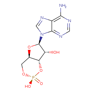
| HET Code: | CMP |
|---|---|
| Formula: | C10H11N5O6P |
| Molecular weight: | 328.198 g/mol |
| DrugBank ID: | DB02527 |
| Buried Surface Area: | 69 % |
| Polar Surface area: | 167.48 Å2 |
| Number of | |
|---|---|
| H-Bond Acceptors: | 10 |
| H-Bond Donors: | 2 |
| Rings: | 4 |
| Aromatic rings: | 2 |
| Anionic atoms: | 1 |
| Cationic atoms: | 0 |
| Rule of Five Violation: | 1 |
| Rotatable Bonds: | 1 |
| X | Y | Z |
|---|---|---|
| 9.447 | 0.0945909 | 40.6153 |
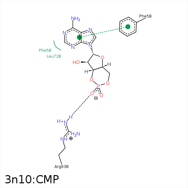


Represent the protein/ligand binding mode, centered on the ligand
Dashed lines represents hydrogen bonds and metal interactions
Green residue labels for amino acids with hydrophobic contacts (green lines) to the ligand
| Ligand | Protein | Interaction | |||
|---|---|---|---|---|---|
| Atom | Atom | Residue | Distance (Å) | Angle (°) | Type |
| C2' | CD2 | PHE- 5 | 3.72 | 0 | Hydrophobic |
| O1P | NH1 | ARG- 63 | 2.88 | 158.56 | H-Bond (Protein Donor) |
| O1P | NH2 | ARG- 63 | 3.29 | 135.44 | H-Bond (Protein Donor) |
| C5' | CD1 | ILE- 74 | 3.48 | 0 | Hydrophobic |
| C4' | SG | CYS- 83 | 4.03 | 0 | Hydrophobic |
| C1' | CB | ALA- 85 | 4.36 | 0 | Hydrophobic |
| O1P | NH1 | ARG- 113 | 3.21 | 128.8 | H-Bond (Protein Donor) |
| C1' | SD | MET- 140 | 4.07 | 0 | Hydrophobic |
| O3' | MN | MN- 183 | 2.4 | 0 | Metal Acceptor |
| O2' | MN | MN- 183 | 2.49 | 0 | Metal Acceptor |
