sc-PDB
An Annotated Database of Druggable Binding Sites from the Protein DataBank
An Annotated Database of Druggable Binding Sites from the Protein DataBank
2.600 Å
X-ray
2010-01-07
| Name: | Putidaredoxin reductase |
|---|---|
| ID: | CAMA_PSEPU |
| AC: | P16640 |
| Organism: | Pseudomonas putida |
| Reign: | Bacteria |
| TaxID: | 303 |
| EC Number: | 1.18.1.5 |
| Chain Name: | Percentage of Residues within binding site |
|---|---|
| A | 100 % |
| B-Factor: | 46.639 |
|---|---|
| Number of residues: | 59 |
| Including | |
| Standard Amino Acids: | 53 |
| Non Standard Amino Acids: | 0 |
| Water Molecules: | 6 |
| Cofactors: | |
| Metals: | |
| Ligandability | Volume (Å3) |
|---|---|
| 0.411 | 2058.750 |
| % Hydrophobic | % Polar |
|---|---|
| 43.77 | 56.23 |
| According to VolSite | |
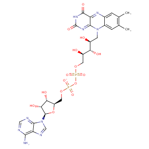
| HET Code: | FAD |
|---|---|
| Formula: | C27H31N9O15P2 |
| Molecular weight: | 783.534 g/mol |
| DrugBank ID: | DB03147 |
| Buried Surface Area: | 72.65 % |
| Polar Surface area: | 381.7 Å2 |
| Number of | |
|---|---|
| H-Bond Acceptors: | 22 |
| H-Bond Donors: | 7 |
| Rings: | 6 |
| Aromatic rings: | 3 |
| Anionic atoms: | 2 |
| Cationic atoms: | 0 |
| Rule of Five Violation: | 3 |
| Rotatable Bonds: | 13 |
| X | Y | Z |
|---|---|---|
| 9.18538 | -6.71838 | -1.23683 |


Represent the protein/ligand binding mode, centered on the ligand
Dashed lines represents hydrogen bonds and metal interactions
Green residue labels for amino acids with hydrophobic contacts (green lines) to the ligand
| Ligand | Protein | Interaction | |||
|---|---|---|---|---|---|
| Atom | Atom | Residue | Distance (Å) | Angle (°) | Type |
| C4' | CB | LEU- 14 | 4.28 | 0 | Hydrophobic |
| O1P | N | ALA- 15 | 2.7 | 143.36 | H-Bond (Protein Donor) |
| O2B | OD2 | ASP- 37 | 2.94 | 167.1 | H-Bond (Ligand Donor) |
| N3A | N | ASP- 37 | 2.86 | 136.67 | H-Bond (Protein Donor) |
| C8M | CG | LEU- 45 | 3.85 | 0 | Hydrophobic |
| C2' | CB | LEU- 45 | 4.38 | 0 | Hydrophobic |
| C3' | CD2 | LEU- 45 | 4.22 | 0 | Hydrophobic |
| C9 | CB | LEU- 45 | 3.66 | 0 | Hydrophobic |
| C7M | CB | LEU- 48 | 4 | 0 | Hydrophobic |
| C6 | CB | SER- 49 | 4.38 | 0 | Hydrophobic |
| C7M | CB | SER- 49 | 3.97 | 0 | Hydrophobic |
| O4 | NZ | LYS- 50 | 2.98 | 161.49 | H-Bond (Protein Donor) |
| N6A | O | VAL- 83 | 2.93 | 168.79 | H-Bond (Ligand Donor) |
| N1A | N | VAL- 83 | 2.95 | 177.86 | H-Bond (Protein Donor) |
| O1A | N | GLY- 112 | 3.31 | 148.64 | H-Bond (Protein Donor) |
| C7M | CG | LEU- 133 | 3.81 | 0 | Hydrophobic |
| O1A | NH2 | ARG- 134 | 3.29 | 149.83 | H-Bond (Protein Donor) |
| O1A | CZ | ARG- 134 | 3.92 | 0 | Ionic (Protein Cationic) |
| C8M | CD | ARG- 134 | 4.16 | 0 | Hydrophobic |
| C6 | CG1 | ILE- 160 | 4.14 | 0 | Hydrophobic |
| C8M | CD1 | ILE- 160 | 4.05 | 0 | Hydrophobic |
| C7 | CD1 | ILE- 160 | 3.66 | 0 | Hydrophobic |
| C7 | CD1 | ILE- 160 | 3.66 | 0 | Hydrophobic |
| O3' | OD1 | ASP- 284 | 2.85 | 140.64 | H-Bond (Ligand Donor) |
| O3' | OD2 | ASP- 284 | 3.49 | 158.67 | H-Bond (Ligand Donor) |
| C5' | CB | ASP- 284 | 4.41 | 0 | Hydrophobic |
| O2P | N | ASP- 284 | 3.06 | 162.14 | H-Bond (Protein Donor) |
| N1 | N | VAL- 302 | 3.45 | 140.65 | H-Bond (Protein Donor) |
| O2 | N | VAL- 302 | 2.94 | 164.52 | H-Bond (Protein Donor) |
| C2' | CG2 | VAL- 302 | 3.69 | 0 | Hydrophobic |
| O1P | O | HOH- 438 | 2.56 | 163.24 | H-Bond (Protein Donor) |
| O3B | O | HOH- 439 | 2.93 | 179.97 | H-Bond (Protein Donor) |
| O2P | O | HOH- 471 | 2.77 | 179.99 | H-Bond (Protein Donor) |
| O1A | O | HOH- 475 | 2.9 | 142.94 | H-Bond (Protein Donor) |
