sc-PDB
An Annotated Database of Druggable Binding Sites from the Protein DataBank
An Annotated Database of Druggable Binding Sites from the Protein DataBank
2.000 Å
X-ray
2009-09-16
| Name: | Secondary alcohol dehydrogenase |
|---|---|
| ID: | Q8KLT9_9NOCA |
| AC: | Q8KLT9 |
| Organism: | Rhodococcus ruber |
| Reign: | Bacteria |
| TaxID: | 1830 |
| EC Number: | / |
| Chain Name: | Percentage of Residues within binding site |
|---|---|
| A | 2 % |
| D | 98 % |
| B-Factor: | 23.516 |
|---|---|
| Number of residues: | 53 |
| Including | |
| Standard Amino Acids: | 49 |
| Non Standard Amino Acids: | 1 |
| Water Molecules: | 3 |
| Cofactors: | |
| Metals: | ZN |
| Ligandability | Volume (Å3) |
|---|---|
| 1.288 | 1036.125 |
| % Hydrophobic | % Polar |
|---|---|
| 51.47 | 48.53 |
| According to VolSite | |
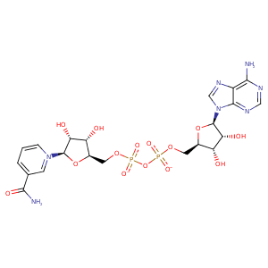
| HET Code: | NAD |
|---|---|
| Formula: | C21H26N7O14P2 |
| Molecular weight: | 662.417 g/mol |
| DrugBank ID: | - |
| Buried Surface Area: | 67.72 % |
| Polar Surface area: | 343.54 Å2 |
| Number of | |
|---|---|
| H-Bond Acceptors: | 18 |
| H-Bond Donors: | 6 |
| Rings: | 5 |
| Aromatic rings: | 3 |
| Anionic atoms: | 2 |
| Cationic atoms: | 1 |
| Rule of Five Violation: | 3 |
| Rotatable Bonds: | 11 |
| X | Y | Z |
|---|---|---|
| 31.2486 | 8.7553 | 59.7385 |


Represent the protein/ligand binding mode, centered on the ligand
Dashed lines represents hydrogen bonds and metal interactions
Green residue labels for amino acids with hydrophobic contacts (green lines) to the ligand
| Ligand | Protein | Interaction | |||
|---|---|---|---|---|---|
| Atom | Atom | Residue | Distance (Å) | Angle (°) | Type |
| C5N | SG | CYS- 38 | 3.88 | 0 | Hydrophobic |
| O1A | ND1 | HIS- 39 | 2.84 | 157.21 | H-Bond (Protein Donor) |
| O1N | N | HIS- 39 | 3.04 | 160.29 | H-Bond (Protein Donor) |
| C5D | CB | HIS- 39 | 4.43 | 0 | Hydrophobic |
| C3D | CB | HIS- 39 | 3.88 | 0 | Hydrophobic |
| C2D | CB | SER- 40 | 4.37 | 0 | Hydrophobic |
| O2D | OG | SER- 40 | 2.65 | 153.79 | H-Bond (Ligand Donor) |
| C4N | CG2 | THR- 157 | 3.54 | 0 | Hydrophobic |
| O2A | N | GLY- 182 | 2.98 | 171.38 | H-Bond (Protein Donor) |
| O2N | N | LEU- 183 | 2.81 | 173.12 | H-Bond (Protein Donor) |
| C5N | CD2 | LEU- 183 | 3.6 | 0 | Hydrophobic |
| O3B | OD1 | ASP- 203 | 2.79 | 154.84 | H-Bond (Ligand Donor) |
| O2B | OD2 | ASP- 203 | 2.71 | 152.15 | H-Bond (Ligand Donor) |
| O2B | OD1 | ASP- 203 | 3.44 | 136.17 | H-Bond (Ligand Donor) |
| N1A | OG | SER- 223 | 2.66 | 158.89 | H-Bond (Protein Donor) |
| C1B | CG2 | VAL- 247 | 4.27 | 0 | Hydrophobic |
| C3N | CG1 | VAL- 269 | 4.35 | 0 | Hydrophobic |
| N7N | O | VAL- 269 | 3.14 | 173.07 | H-Bond (Ligand Donor) |
| O3D | N | ILE- 271 | 2.96 | 173.61 | H-Bond (Protein Donor) |
| N7N | O | PRO- 293 | 3.08 | 166.56 | H-Bond (Ligand Donor) |
| O7N | N | TRP- 295 | 2.73 | 159.03 | H-Bond (Protein Donor) |
| O1N | NH1 | ARG- 340 | 2.88 | 149.04 | H-Bond (Protein Donor) |
| O1N | CZ | ARG- 340 | 3.92 | 0 | Ionic (Protein Cationic) |
| O2N | O | HOH- 354 | 2.82 | 166.3 | H-Bond (Protein Donor) |
| O2A | O | HOH- 416 | 2.57 | 176.62 | H-Bond (Protein Donor) |
| O2A | O | HOH- 453 | 2.75 | 162.96 | H-Bond (Protein Donor) |
