sc-PDB
An Annotated Database of Druggable Binding Sites from the Protein DataBank
An Annotated Database of Druggable Binding Sites from the Protein DataBank
2.400 Å
X-ray
2009-05-07
| Name: | UDP-galactopyranose mutase |
|---|---|
| ID: | Q9RYF1_DEIRA |
| AC: | Q9RYF1 |
| Organism: | Deinococcus radiodurans |
| Reign: | Bacteria |
| TaxID: | 243230 |
| EC Number: | / |
| Chain Name: | Percentage of Residues within binding site |
|---|---|
| B | 100 % |
| B-Factor: | 47.215 |
|---|---|
| Number of residues: | 59 |
| Including | |
| Standard Amino Acids: | 56 |
| Non Standard Amino Acids: | 1 |
| Water Molecules: | 2 |
| Cofactors: | |
| Metals: | |
| Ligandability | Volume (Å3) |
|---|---|
| 0.846 | 1238.625 |
| % Hydrophobic | % Polar |
|---|---|
| 45.23 | 54.77 |
| According to VolSite | |
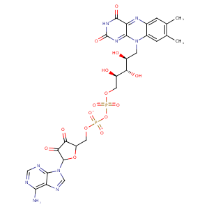
| HET Code: | FDA |
|---|---|
| Formula: | C27H33N9O15P2 |
| Molecular weight: | 785.550 g/mol |
| DrugBank ID: | - |
| Buried Surface Area: | 71.86 % |
| Polar Surface area: | 381.04 Å2 |
| Number of | |
|---|---|
| H-Bond Acceptors: | 21 |
| H-Bond Donors: | 9 |
| Rings: | 6 |
| Aromatic rings: | 3 |
| Anionic atoms: | 2 |
| Cationic atoms: | 0 |
| Rule of Five Violation: | 3 |
| Rotatable Bonds: | 13 |
| X | Y | Z |
|---|---|---|
| -2.58179 | -58.4727 | -80.4976 |
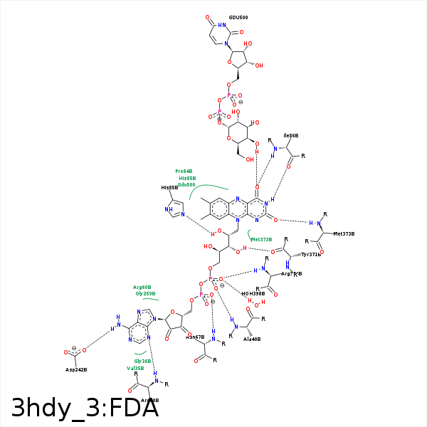


Represent the protein/ligand binding mode, centered on the ligand
Dashed lines represents hydrogen bonds and metal interactions
Green residue labels for amino acids with hydrophobic contacts (green lines) to the ligand
| Ligand | Protein | Interaction | |||
|---|---|---|---|---|---|
| Atom | Atom | Residue | Distance (Å) | Angle (°) | Type |
| O2P | N | ALA- 40 | 3.14 | 152.65 | H-Bond (Protein Donor) |
| N3A | N | ARG- 60 | 3.21 | 154.43 | H-Bond (Protein Donor) |
| O1A | N | ASN- 67 | 3.19 | 174.92 | H-Bond (Protein Donor) |
| O2' | ND2 | ASN- 67 | 3.42 | 143.89 | H-Bond (Protein Donor) |
| C2' | CB | ASN- 67 | 4.49 | 0 | Hydrophobic |
| O2' | NE2 | HIS- 85 | 3.03 | 152.7 | H-Bond (Ligand Donor) |
| DuAr | DuAr | HIS- 85 | 4 | 0 | Aromatic Face/Face |
| N3 | O | ILE- 86 | 3.01 | 160.47 | H-Bond (Ligand Donor) |
| O4 | N | ILE- 86 | 3.1 | 172.53 | H-Bond (Protein Donor) |
| N6A | OD1 | ASP- 242 | 3.2 | 161.02 | H-Bond (Ligand Donor) |
| N1A | N | TYR- 243 | 3.38 | 156.36 | H-Bond (Protein Donor) |
| C7M | CD1 | LEU- 277 | 3.59 | 0 | Hydrophobic |
| C7M | CZ | PHE- 279 | 4.03 | 0 | Hydrophobic |
| C8M | CB | TYR- 334 | 3.71 | 0 | Hydrophobic |
| C3' | CD | ARG- 364 | 4.38 | 0 | Hydrophobic |
| C5' | CB | ARG- 364 | 4.27 | 0 | Hydrophobic |
| O1P | N | ARG- 364 | 3.06 | 159.45 | H-Bond (Protein Donor) |
| C5B | CD1 | LEU- 365 | 4.29 | 0 | Hydrophobic |
| O3' | O | TYR- 371 | 2.94 | 151.86 | H-Bond (Ligand Donor) |
| O2 | N | MET- 373 | 2.85 | 163.55 | H-Bond (Protein Donor) |
| C2' | CG | MET- 373 | 4.12 | 0 | Hydrophobic |
| C5' | CG2 | VAL- 376 | 3.7 | 0 | Hydrophobic |
| O1P | O | HOH- 398 | 2.93 | 179.95 | H-Bond (Protein Donor) |
