sc-PDB
An Annotated Database of Druggable Binding Sites from the Protein DataBank
An Annotated Database of Druggable Binding Sites from the Protein DataBank
2.300 Å
X-ray
2009-03-31
| Name: | Cyclohexanone monooxygenase |
|---|---|
| ID: | C0STX7_9NOCA |
| AC: | C0STX7 |
| Organism: | Rhodococcus sp. HI-31 |
| Reign: | Bacteria |
| TaxID: | 638919 |
| EC Number: | / |
| Chain Name: | Percentage of Residues within binding site |
|---|---|
| A | 100 % |
| B-Factor: | 41.261 |
|---|---|
| Number of residues: | 62 |
| Including | |
| Standard Amino Acids: | 56 |
| Non Standard Amino Acids: | 2 |
| Water Molecules: | 4 |
| Cofactors: | NAP NAP |
| Metals: | |
| Ligandability | Volume (Å3) |
|---|---|
| 0.998 | 428.625 |
| % Hydrophobic | % Polar |
|---|---|
| 46.46 | 53.54 |
| According to VolSite | |
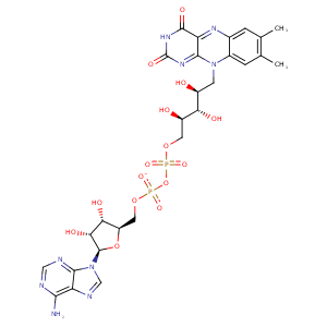
| HET Code: | FAD |
|---|---|
| Formula: | C27H31N9O15P2 |
| Molecular weight: | 783.534 g/mol |
| DrugBank ID: | DB03147 |
| Buried Surface Area: | 77.46 % |
| Polar Surface area: | 381.7 Å2 |
| Number of | |
|---|---|
| H-Bond Acceptors: | 22 |
| H-Bond Donors: | 7 |
| Rings: | 6 |
| Aromatic rings: | 3 |
| Anionic atoms: | 2 |
| Cationic atoms: | 0 |
| Rule of Five Violation: | 3 |
| Rotatable Bonds: | 13 |
| X | Y | Z |
|---|---|---|
| -12.7224 | 3.25419 | 6.67628 |


Represent the protein/ligand binding mode, centered on the ligand
Dashed lines represents hydrogen bonds and metal interactions
Green residue labels for amino acids with hydrophobic contacts (green lines) to the ligand
| Ligand | Protein | Interaction | |||
|---|---|---|---|---|---|
| Atom | Atom | Residue | Distance (Å) | Angle (°) | Type |
| C4' | CD2 | PHE- 18 | 4.06 | 0 | Hydrophobic |
| O1P | N | GLY- 19 | 2.78 | 156.79 | H-Bond (Protein Donor) |
| O3B | OD2 | ASP- 39 | 2.6 | 157.8 | H-Bond (Ligand Donor) |
| O3B | OD1 | ASP- 39 | 3.27 | 137.51 | H-Bond (Ligand Donor) |
| O2B | OD1 | ASP- 39 | 2.77 | 156.22 | H-Bond (Ligand Donor) |
| N3A | N | LYS- 40 | 3.49 | 147.72 | H-Bond (Protein Donor) |
| O2A | N | THR- 47 | 2.87 | 166.99 | H-Bond (Protein Donor) |
| O2A | OG1 | THR- 47 | 2.96 | 173.89 | H-Bond (Protein Donor) |
| C2' | CG2 | THR- 47 | 4.31 | 0 | Hydrophobic |
| C7 | CG2 | THR- 47 | 3.88 | 0 | Hydrophobic |
| C8 | CG2 | THR- 47 | 3.33 | 0 | Hydrophobic |
| O2' | OG1 | THR- 47 | 3.12 | 155.91 | H-Bond (Ligand Donor) |
| O4' | OG1 | THR- 47 | 3.33 | 160.08 | H-Bond (Ligand Donor) |
| C6 | CH2 | TRP- 48 | 3.36 | 0 | Hydrophobic |
| O3B | NE1 | TRP- 50 | 3.18 | 135 | H-Bond (Protein Donor) |
| O2B | NE1 | TRP- 50 | 2.96 | 139.18 | H-Bond (Protein Donor) |
| C7M | CB | ASN- 51 | 4.27 | 0 | Hydrophobic |
| C7M | CE2 | TYR- 53 | 3.91 | 0 | Hydrophobic |
| O4 | N | ASP- 59 | 2.7 | 131.51 | H-Bond (Protein Donor) |
| N3 | OG1 | THR- 60 | 2.7 | 177.24 | H-Bond (Ligand Donor) |
| O4 | N | THR- 60 | 3.1 | 140.84 | H-Bond (Protein Donor) |
| O3' | OH | TYR- 65 | 2.66 | 157.71 | H-Bond (Protein Donor) |
| N6A | O | VAL- 112 | 2.97 | 174.5 | H-Bond (Ligand Donor) |
| N1A | N | VAL- 112 | 2.9 | 146.23 | H-Bond (Protein Donor) |
| C1' | CD1 | LEU- 146 | 4.25 | 0 | Hydrophobic |
| C8M | CZ | PHE- 382 | 3.98 | 0 | Hydrophobic |
| C5' | CD1 | LEU- 428 | 4.47 | 0 | Hydrophobic |
| O2 | N | LEU- 437 | 2.75 | 170.9 | H-Bond (Protein Donor) |
| C3' | CD1 | LEU- 437 | 3.8 | 0 | Hydrophobic |
| C5' | CD1 | ILE- 441 | 4.13 | 0 | Hydrophobic |
| C7M | C4N | NAP- 542 | 3.48 | 0 | Hydrophobic |
| O2P | O | HOH- 545 | 2.58 | 153.9 | H-Bond (Protein Donor) |
| O1P | O | HOH- 546 | 2.72 | 165.38 | H-Bond (Protein Donor) |
