sc-PDB
An Annotated Database of Druggable Binding Sites from the Protein DataBank
An Annotated Database of Druggable Binding Sites from the Protein DataBank
1.980 Å
X-ray
2009-03-18
| Name: | Ribokinase |
|---|---|
| ID: | P71913_MYCTU |
| AC: | P71913 |
| Organism: | Mycobacterium tuberculosis |
| Reign: | Bacteria |
| TaxID: | 83332 |
| EC Number: | / |
| Chain Name: | Percentage of Residues within binding site |
|---|---|
| B | 100 % |
| B-Factor: | 27.601 |
|---|---|
| Number of residues: | 36 |
| Including | |
| Standard Amino Acids: | 32 |
| Non Standard Amino Acids: | 0 |
| Water Molecules: | 4 |
| Cofactors: | |
| Metals: | |
| Ligandability | Volume (Å3) |
|---|---|
| 0.982 | 253.125 |
| % Hydrophobic | % Polar |
|---|---|
| 57.33 | 42.67 |
| According to VolSite | |
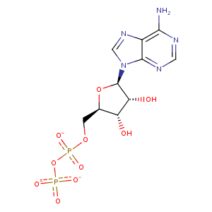
| HET Code: | ADP |
|---|---|
| Formula: | C10H12N5O10P2 |
| Molecular weight: | 424.177 g/mol |
| DrugBank ID: | - |
| Buried Surface Area: | 64.69 % |
| Polar Surface area: | 260.7 Å2 |
| Number of | |
|---|---|
| H-Bond Acceptors: | 14 |
| H-Bond Donors: | 3 |
| Rings: | 3 |
| Aromatic rings: | 2 |
| Anionic atoms: | 3 |
| Cationic atoms: | 0 |
| Rule of Five Violation: | 1 |
| Rotatable Bonds: | 6 |
| X | Y | Z |
|---|---|---|
| -8.06111 | -1.52219 | 31.7878 |
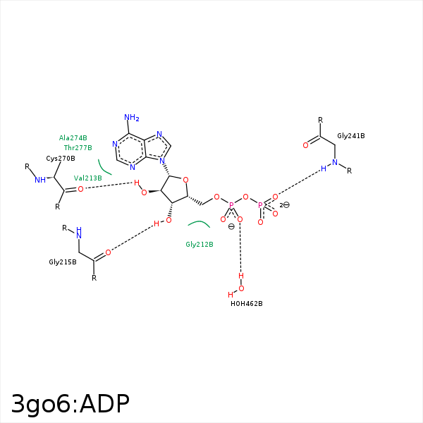


Represent the protein/ligand binding mode, centered on the ligand
Dashed lines represents hydrogen bonds and metal interactions
Green residue labels for amino acids with hydrophobic contacts (green lines) to the ligand
| Ligand | Protein | Interaction | |||
|---|---|---|---|---|---|
| Atom | Atom | Residue | Distance (Å) | Angle (°) | Type |
| C5' | CB | THR- 210 | 4.03 | 0 | Hydrophobic |
| C3' | CB | THR- 210 | 4.34 | 0 | Hydrophobic |
| O3' | O | GLY- 215 | 2.63 | 153.47 | H-Bond (Ligand Donor) |
| C2' | CB | ALA- 229 | 4.37 | 0 | Hydrophobic |
| O1B | OG1 | THR- 237 | 3.4 | 158.76 | H-Bond (Protein Donor) |
| O3B | N | GLY- 241 | 2.87 | 160.56 | H-Bond (Protein Donor) |
| C4' | CD1 | PHE- 244 | 4.14 | 0 | Hydrophobic |
| O2' | O | CYS- 270 | 2.89 | 126.96 | H-Bond (Ligand Donor) |
| O5' | O | HOH- 462 | 3.46 | 130.46 | H-Bond (Protein Donor) |
