sc-PDB
An Annotated Database of Druggable Binding Sites from the Protein DataBank
An Annotated Database of Druggable Binding Sites from the Protein DataBank
2.500 Å
X-ray
2008-12-10
| Name: | Pyridoxal kinase |
|---|---|
| ID: | PDXK_HUMAN |
| AC: | O00764 |
| Organism: | Homo sapiens |
| Reign: | Eukaryota |
| TaxID: | 9606 |
| EC Number: | 2.7.1.35 |
| Chain Name: | Percentage of Residues within binding site |
|---|---|
| A | 100 % |
| B-Factor: | 43.699 |
|---|---|
| Number of residues: | 20 |
| Including | |
| Standard Amino Acids: | 19 |
| Non Standard Amino Acids: | 1 |
| Water Molecules: | 0 |
| Cofactors: | ATP |
| Metals: | |
| Ligandability | Volume (Å3) |
|---|---|
| 0.869 | 759.375 |
| % Hydrophobic | % Polar |
|---|---|
| 52.89 | 47.11 |
| According to VolSite | |
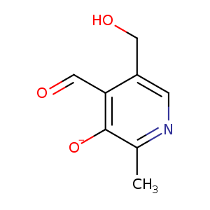
| HET Code: | PXL |
|---|---|
| Formula: | C8H9NO3 |
| Molecular weight: | 167.162 g/mol |
| DrugBank ID: | DB00147 |
| Buried Surface Area: | 43.77 % |
| Polar Surface area: | 70.42 Å2 |
| Number of | |
|---|---|
| H-Bond Acceptors: | 4 |
| H-Bond Donors: | 2 |
| Rings: | 1 |
| Aromatic rings: | 1 |
| Anionic atoms: | 0 |
| Cationic atoms: | 0 |
| Rule of Five Violation: | 0 |
| Rotatable Bonds: | 2 |
| X | Y | Z |
|---|---|---|
| 30.1197 | 6.32758 | 38.5318 |
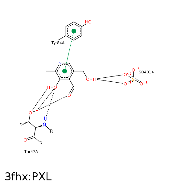


Represent the protein/ligand binding mode, centered on the ligand
Dashed lines represents hydrogen bonds and metal interactions
Green residue labels for amino acids with hydrophobic contacts (green lines) to the ligand
| Ligand | Protein | Interaction | |||
|---|---|---|---|---|---|
| Atom | Atom | Residue | Distance (Å) | Angle (°) | Type |
| N1 | OG | SER- 12 | 3.33 | 149.23 | H-Bond (Protein Donor) |
| C2A | CG2 | VAL- 41 | 4.46 | 0 | Hydrophobic |
| C3 | CB | HIS- 46 | 3.93 | 0 | Hydrophobic |
| C3 | CG2 | THR- 47 | 4.15 | 0 | Hydrophobic |
| C2A | CG2 | THR- 47 | 4.38 | 0 | Hydrophobic |
| O4A | OG1 | THR- 47 | 3.03 | 138.6 | H-Bond (Protein Donor) |
| O3 | N | THR- 47 | 2.61 | 174.32 | H-Bond (Protein Donor) |
| O3 | OG1 | THR- 47 | 3.04 | 143.67 | H-Bond (Protein Donor) |
| C5A | CG1 | VAL- 231 | 4.41 | 0 | Hydrophobic |
