sc-PDB
An Annotated Database of Druggable Binding Sites from the Protein DataBank
An Annotated Database of Druggable Binding Sites from the Protein DataBank
2.500 Å
X-ray
2008-10-07
| Name: | Glutamate dehydrogenase 1, mitochondrial |
|---|---|
| ID: | DHE3_BOVIN |
| AC: | P00366 |
| Organism: | Bos taurus |
| Reign: | Eukaryota |
| TaxID: | 9913 |
| EC Number: | 1.4.1.3 |
| Chain Name: | Percentage of Residues within binding site |
|---|---|
| D | 24 % |
| F | 76 % |
| B-Factor: | 39.621 |
|---|---|
| Number of residues: | 22 |
| Including | |
| Standard Amino Acids: | 21 |
| Non Standard Amino Acids: | 1 |
| Water Molecules: | 0 |
| Cofactors: | |
| Metals: | |
| Ligandability | Volume (Å3) |
|---|---|
| 1.055 | 1491.750 |
| % Hydrophobic | % Polar |
|---|---|
| 39.82 | 60.18 |
| According to VolSite | |
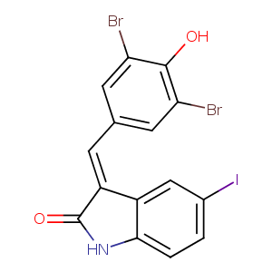
| HET Code: | GWD |
|---|---|
| Formula: | C15H7Br2INO2 |
| Molecular weight: | 519.934 g/mol |
| DrugBank ID: | - |
| Buried Surface Area: | 37.36 % |
| Polar Surface area: | 52.16 Å2 |
| Number of | |
|---|---|
| H-Bond Acceptors: | 2 |
| H-Bond Donors: | 1 |
| Rings: | 3 |
| Aromatic rings: | 2 |
| Anionic atoms: | 1 |
| Cationic atoms: | 0 |
| Rule of Five Violation: | 1 |
| Rotatable Bonds: | 1 |
| X | Y | Z |
|---|---|---|
| 36.7762 | 36.9495 | 140.921 |
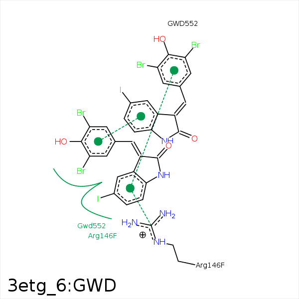


Represent the protein/ligand binding mode, centered on the ligand
Dashed lines represents hydrogen bonds and metal interactions
Green residue labels for amino acids with hydrophobic contacts (green lines) to the ligand
| Ligand | Protein | Interaction | |||
|---|---|---|---|---|---|
| Atom | Atom | Residue | Distance (Å) | Angle (°) | Type |
| BRAD | CB | LYS- 143 | 4.45 | 0 | Hydrophobic |
| CAI | CB | ARG- 146 | 4.25 | 0 | Hydrophobic |
| IAE | CD | ARG- 146 | 4.24 | 0 | Hydrophobic |
| CAK | CD | ARG- 146 | 3.24 | 0 | Hydrophobic |
| DuAr | CZ | ARG- 146 | 3.75 | 20.25 | Pi/Cation |
| OAB | NH1 | ARG- 147 | 3.48 | 148.5 | H-Bond (Protein Donor) |
| BRAC | CD | ARG- 147 | 3.65 | 0 | Hydrophobic |
| BRAC | CG | MET- 150 | 3.9 | 0 | Hydrophobic |
| CAH | CG | MET- 150 | 3.45 | 0 | Hydrophobic |
| BRAC | CB | SER- 185 | 4.34 | 0 | Hydrophobic |
