sc-PDB
An Annotated Database of Druggable Binding Sites from the Protein DataBank
An Annotated Database of Druggable Binding Sites from the Protein DataBank
2.300 Å
X-ray
2008-06-23
| Name: | Purine nucleoside phosphorylase |
|---|---|
| ID: | Q9BMI9_SCHMA |
| AC: | Q9BMI9 |
| Organism: | Schistosoma mansoni |
| Reign: | Eukaryota |
| TaxID: | 6183 |
| EC Number: | / |
| Chain Name: | Percentage of Residues within binding site |
|---|---|
| A | 97 % |
| B | 3 % |
| B-Factor: | 35.477 |
|---|---|
| Number of residues: | 31 |
| Including | |
| Standard Amino Acids: | 30 |
| Non Standard Amino Acids: | 0 |
| Water Molecules: | 1 |
| Cofactors: | |
| Metals: | |
| Ligandability | Volume (Å3) |
|---|---|
| 1.093 | 448.875 |
| % Hydrophobic | % Polar |
|---|---|
| 57.89 | 42.11 |
| According to VolSite | |
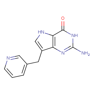
| HET Code: | BC3 |
|---|---|
| Formula: | C12H11N5O |
| Molecular weight: | 241.249 g/mol |
| DrugBank ID: | DB02568 |
| Buried Surface Area: | 69.94 % |
| Polar Surface area: | 96.16 Å2 |
| Number of | |
|---|---|
| H-Bond Acceptors: | 4 |
| H-Bond Donors: | 3 |
| Rings: | 3 |
| Aromatic rings: | 2 |
| Anionic atoms: | 0 |
| Cationic atoms: | 0 |
| Rule of Five Violation: | 0 |
| Rotatable Bonds: | 2 |
| X | Y | Z |
|---|---|---|
| -18.0807 | -2.59344 | 28.5311 |
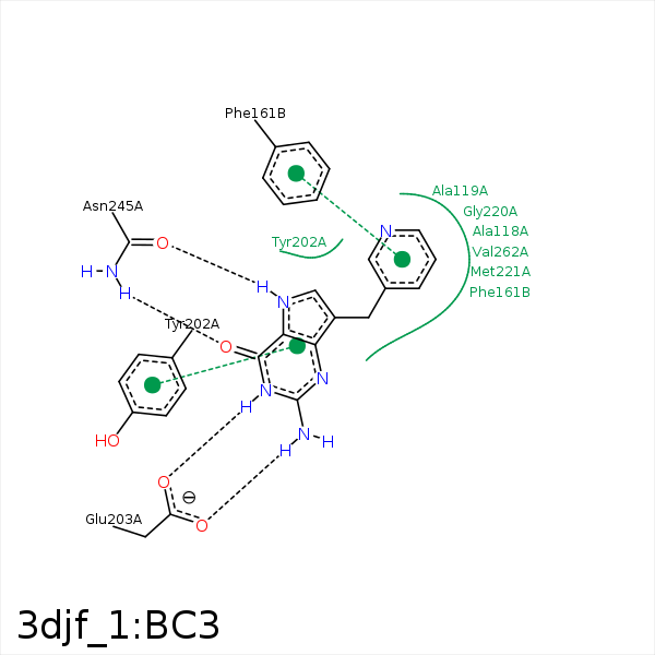


Represent the protein/ligand binding mode, centered on the ligand
Dashed lines represents hydrogen bonds and metal interactions
Green residue labels for amino acids with hydrophobic contacts (green lines) to the ligand
| Ligand | Protein | Interaction | |||
|---|---|---|---|---|---|
| Atom | Atom | Residue | Distance (Å) | Angle (°) | Type |
| C06 | CB | ALA- 118 | 4.13 | 0 | Hydrophobic |
| N01 | OE2 | GLU- 203 | 2.68 | 173.55 | H-Bond (Ligand Donor) |
| N18 | OE1 | GLU- 203 | 2.8 | 170.44 | H-Bond (Ligand Donor) |
| C08 | CE | MET- 221 | 3.69 | 0 | Hydrophobic |
| N14 | OD1 | ASN- 245 | 2.85 | 172.46 | H-Bond (Ligand Donor) |
| O17 | ND2 | ASN- 245 | 2.97 | 177.6 | H-Bond (Protein Donor) |
