sc-PDB
An Annotated Database of Druggable Binding Sites from the Protein DataBank
An Annotated Database of Druggable Binding Sites from the Protein DataBank
2.100 Å
X-ray
2008-02-07
| Name: | Octopine dehydrogenase |
|---|---|
| ID: | OCDH_PECMA |
| AC: | Q9BHM6 |
| Organism: | Pecten maximus |
| Reign: | Eukaryota |
| TaxID: | 6579 |
| EC Number: | / |
| Chain Name: | Percentage of Residues within binding site |
|---|---|
| A | 100 % |
| B-Factor: | 15.947 |
|---|---|
| Number of residues: | 35 |
| Including | |
| Standard Amino Acids: | 31 |
| Non Standard Amino Acids: | 0 |
| Water Molecules: | 4 |
| Cofactors: | |
| Metals: | |
| Ligandability | Volume (Å3) |
|---|---|
| 0.066 | 263.250 |
| % Hydrophobic | % Polar |
|---|---|
| 42.31 | 57.69 |
| According to VolSite | |
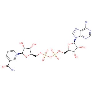
| HET Code: | NAD |
|---|---|
| Formula: | C21H26N7O14P2 |
| Molecular weight: | 662.417 g/mol |
| DrugBank ID: | - |
| Buried Surface Area: | 38.3 % |
| Polar Surface area: | 343.54 Å2 |
| Number of | |
|---|---|
| H-Bond Acceptors: | 18 |
| H-Bond Donors: | 6 |
| Rings: | 5 |
| Aromatic rings: | 3 |
| Anionic atoms: | 2 |
| Cationic atoms: | 1 |
| Rule of Five Violation: | 3 |
| Rotatable Bonds: | 11 |
| X | Y | Z |
|---|---|---|
| 88.9167 | 34.5572 | 53.5906 |
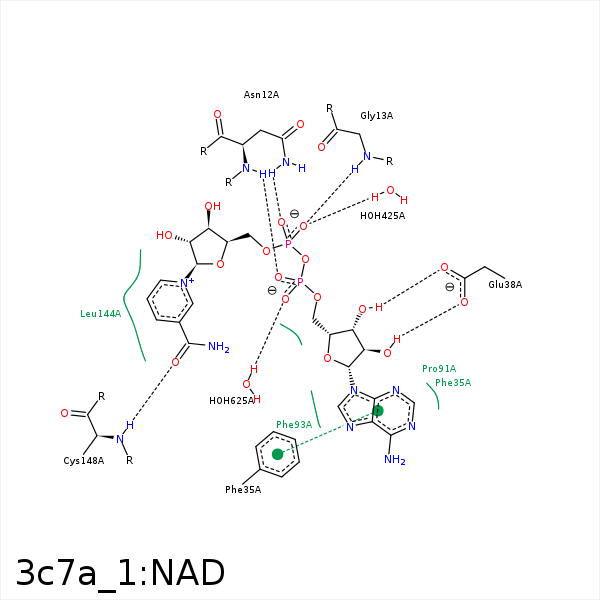


Represent the protein/ligand binding mode, centered on the ligand
Dashed lines represents hydrogen bonds and metal interactions
Green residue labels for amino acids with hydrophobic contacts (green lines) to the ligand
| Ligand | Protein | Interaction | |||
|---|---|---|---|---|---|
| Atom | Atom | Residue | Distance (Å) | Angle (°) | Type |
| O2A | N | ASN- 12 | 3.2 | 170.91 | H-Bond (Protein Donor) |
| O1N | ND2 | ASN- 12 | 3.16 | 152.34 | H-Bond (Protein Donor) |
| O2N | N | GLY- 13 | 2.94 | 161.83 | H-Bond (Protein Donor) |
| C2B | CD2 | PHE- 35 | 4.12 | 0 | Hydrophobic |
| O3B | OE2 | GLU- 38 | 3.13 | 165.93 | H-Bond (Ligand Donor) |
| O2B | OE1 | GLU- 38 | 3.04 | 162.96 | H-Bond (Ligand Donor) |
| C5N | CB | LEU- 144 | 3.96 | 0 | Hydrophobic |
| C3N | CD2 | LEU- 144 | 3.64 | 0 | Hydrophobic |
| O7N | N | CYS- 148 | 3.28 | 161.37 | H-Bond (Protein Donor) |
| O2N | O | HOH- 425 | 2.88 | 154.01 | H-Bond (Protein Donor) |
| O1A | O | HOH- 625 | 3.05 | 179.96 | H-Bond (Protein Donor) |
