sc-PDB
An Annotated Database of Druggable Binding Sites from the Protein DataBank
An Annotated Database of Druggable Binding Sites from the Protein DataBank
3.000 Å
X-ray
2008-01-28
| Name: | 2-phospho-L-lactate transferase |
|---|---|
| ID: | COFD_METMA |
| AC: | Q8PVT6 |
| Organism: | Methanosarcina mazei |
| Reign: | Archaea |
| TaxID: | 192952 |
| EC Number: | / |
| Chain Name: | Percentage of Residues within binding site |
|---|---|
| B | 100 % |
| B-Factor: | 77.923 |
|---|---|
| Number of residues: | 21 |
| Including | |
| Standard Amino Acids: | 21 |
| Non Standard Amino Acids: | 0 |
| Water Molecules: | 0 |
| Cofactors: | |
| Metals: | |
| Ligandability | Volume (Å3) |
|---|---|
| 0.810 | 594.000 |
| % Hydrophobic | % Polar |
|---|---|
| 53.98 | 46.02 |
| According to VolSite | |
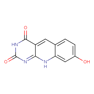
| HET Code: | FO1 |
|---|---|
| Formula: | C16H17N3O7 |
| Molecular weight: | 363.322 g/mol |
| DrugBank ID: | - |
| Buried Surface Area: | 59.98 % |
| Polar Surface area: | 162.91 Å2 |
| Number of | |
|---|---|
| H-Bond Acceptors: | 9 |
| H-Bond Donors: | 6 |
| Rings: | 3 |
| Aromatic rings: | 1 |
| Anionic atoms: | 0 |
| Cationic atoms: | 0 |
| Rule of Five Violation: | 1 |
| Rotatable Bonds: | 5 |
| X | Y | Z |
|---|---|---|
| 127.453 | 10.8607 | -2.28412 |
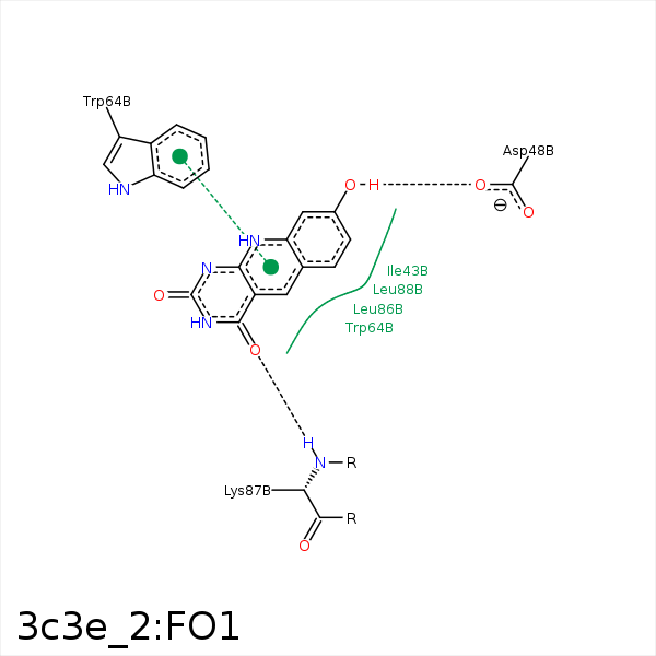


Represent the protein/ligand binding mode, centered on the ligand
Dashed lines represents hydrogen bonds and metal interactions
Green residue labels for amino acids with hydrophobic contacts (green lines) to the ligand
| Ligand | Protein | Interaction | |||
|---|---|---|---|---|---|
| Atom | Atom | Residue | Distance (Å) | Angle (°) | Type |
| C7 | CG2 | ILE- 43 | 3.83 | 0 | Hydrophobic |
| C11 | CB | PRO- 45 | 3.68 | 0 | Hydrophobic |
| O10 | OD1 | ASP- 48 | 3.23 | 140.53 | H-Bond (Ligand Donor) |
| C5 | CE3 | TRP- 64 | 3.49 | 0 | Hydrophobic |
| C12 | CB | TRP- 64 | 3.94 | 0 | Hydrophobic |
| C6 | CD1 | LEU- 86 | 3.59 | 0 | Hydrophobic |
| O2 | N | LYS- 87 | 3.01 | 146.1 | H-Bond (Protein Donor) |
| C6 | CD2 | LEU- 88 | 3.52 | 0 | Hydrophobic |
