sc-PDB
An Annotated Database of Druggable Binding Sites from the Protein DataBank
An Annotated Database of Druggable Binding Sites from the Protein DataBank
2.400 Å
X-ray
2008-01-28
| Name: | Phosphoglycerate kinase 1 |
|---|---|
| ID: | PGK1_HUMAN |
| AC: | P00558 |
| Organism: | Homo sapiens |
| Reign: | Eukaryota |
| TaxID: | 9606 |
| EC Number: | 2.7.2.3 |
| Chain Name: | Percentage of Residues within binding site |
|---|---|
| A | 100 % |
| B-Factor: | 27.826 |
|---|---|
| Number of residues: | 28 |
| Including | |
| Standard Amino Acids: | 27 |
| Non Standard Amino Acids: | 1 |
| Water Molecules: | 0 |
| Cofactors: | |
| Metals: | MG |
| Ligandability | Volume (Å3) |
|---|---|
| 0.770 | 290.250 |
| % Hydrophobic | % Polar |
|---|---|
| 68.60 | 31.40 |
| According to VolSite | |
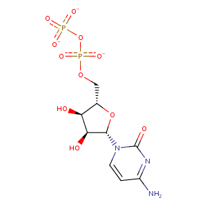
| HET Code: | CDP |
|---|---|
| Formula: | C9H12N3O11P2 |
| Molecular weight: | 400.153 g/mol |
| DrugBank ID: | DB04555 |
| Buried Surface Area: | 50.69 % |
| Polar Surface area: | 249.77 Å2 |
| Number of | |
|---|---|
| H-Bond Acceptors: | 13 |
| H-Bond Donors: | 3 |
| Rings: | 2 |
| Aromatic rings: | 0 |
| Anionic atoms: | 3 |
| Cationic atoms: | 0 |
| Rule of Five Violation: | 1 |
| Rotatable Bonds: | 6 |
| X | Y | Z |
|---|---|---|
| 12.1752 | -0.9662 | 34.5753 |
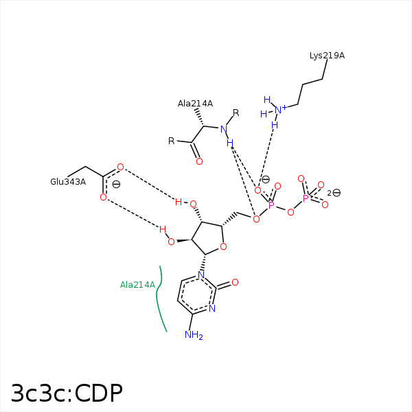


Represent the protein/ligand binding mode, centered on the ligand
Dashed lines represents hydrogen bonds and metal interactions
Green residue labels for amino acids with hydrophobic contacts (green lines) to the ligand
| Ligand | Protein | Interaction | |||
|---|---|---|---|---|---|
| Atom | Atom | Residue | Distance (Å) | Angle (°) | Type |
| O1A | N | ALA- 214 | 2.93 | 148.53 | H-Bond (Protein Donor) |
| O5' | N | ALA- 214 | 3.13 | 138.75 | H-Bond (Protein Donor) |
| C5' | CB | ALA- 214 | 4.35 | 0 | Hydrophobic |
| O2B | NZ | LYS- 219 | 3.77 | 0 | Ionic (Protein Cationic) |
| O1A | NZ | LYS- 219 | 2.76 | 0 | Ionic (Protein Cationic) |
| O1A | NZ | LYS- 219 | 2.76 | 129.17 | H-Bond (Protein Donor) |
| O2 | N | GLY- 238 | 3.21 | 127.11 | H-Bond (Protein Donor) |
| C1' | CG | PRO- 338 | 3.76 | 0 | Hydrophobic |
| O3' | OE2 | GLU- 343 | 2.73 | 166.34 | H-Bond (Ligand Donor) |
| O2' | OE1 | GLU- 343 | 2.89 | 159.97 | H-Bond (Ligand Donor) |
