sc-PDB
An Annotated Database of Druggable Binding Sites from the Protein DataBank
An Annotated Database of Druggable Binding Sites from the Protein DataBank
2.550 Å
X-ray
2009-12-28
| Name: | Tyrosine-protein kinase Lck |
|---|---|
| ID: | LCK_HUMAN |
| AC: | P06239 |
| Organism: | Homo sapiens |
| Reign: | Eukaryota |
| TaxID: | 9606 |
| EC Number: | 2.7.10.2 |
| Chain Name: | Percentage of Residues within binding site |
|---|---|
| A | 100 % |
| B-Factor: | 5.820 |
|---|---|
| Number of residues: | 27 |
| Including | |
| Standard Amino Acids: | 27 |
| Non Standard Amino Acids: | 0 |
| Water Molecules: | 0 |
| Cofactors: | |
| Metals: | |
| Ligandability | Volume (Å3) |
|---|---|
| 1.234 | 472.500 |
| % Hydrophobic | % Polar |
|---|---|
| 57.86 | 42.14 |
| According to VolSite | |
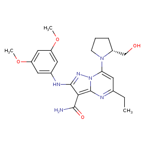
| HET Code: | KSH |
|---|---|
| Formula: | C22H28N6O4 |
| Molecular weight: | 440.496 g/mol |
| DrugBank ID: | - |
| Buried Surface Area: | 37.91 % |
| Polar Surface area: | 127.24 Å2 |
| Number of | |
|---|---|
| H-Bond Acceptors: | 8 |
| H-Bond Donors: | 3 |
| Rings: | 4 |
| Aromatic rings: | 3 |
| Anionic atoms: | 0 |
| Cationic atoms: | 0 |
| Rule of Five Violation: | 0 |
| Rotatable Bonds: | 8 |
| X | Y | Z |
|---|---|---|
| 7.11084 | 1.31637 | -8.25541 |
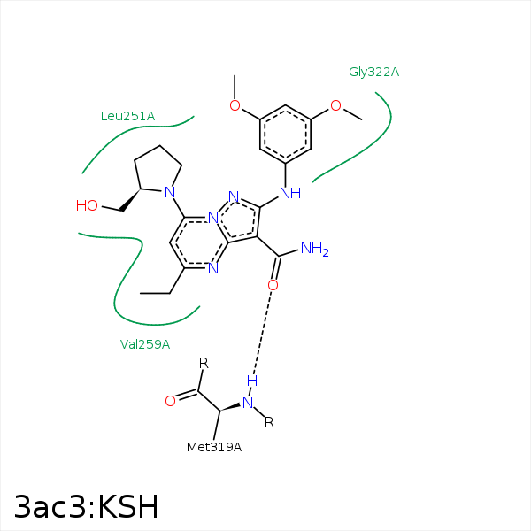


Represent the protein/ligand binding mode, centered on the ligand
Dashed lines represents hydrogen bonds and metal interactions
Green residue labels for amino acids with hydrophobic contacts (green lines) to the ligand
| Ligand | Protein | Interaction | |||
|---|---|---|---|---|---|
| Atom | Atom | Residue | Distance (Å) | Angle (°) | Type |
| C19 | CD2 | LEU- 251 | 4.48 | 0 | Hydrophobic |
| C32 | CD2 | LEU- 251 | 4.4 | 0 | Hydrophobic |
| C18 | CD1 | LEU- 251 | 3.6 | 0 | Hydrophobic |
| O11 | N | MET- 319 | 2.77 | 141.8 | H-Bond (Protein Donor) |
| C53 | CB | SER- 323 | 3.68 | 0 | Hydrophobic |
