sc-PDB
An Annotated Database of Druggable Binding Sites from the Protein DataBank
An Annotated Database of Druggable Binding Sites from the Protein DataBank
1.900 Å
X-ray
2009-08-17
| Name: | Elongation factor P--(R)-beta-lysine ligase |
|---|---|
| ID: | EPMA_ECOLI |
| AC: | P0A8N7 |
| Organism: | Escherichia coli |
| Reign: | Bacteria |
| TaxID: | 83333 |
| EC Number: | / |
| Chain Name: | Percentage of Residues within binding site |
|---|---|
| C | 100 % |
| B-Factor: | 20.572 |
|---|---|
| Number of residues: | 47 |
| Including | |
| Standard Amino Acids: | 44 |
| Non Standard Amino Acids: | 0 |
| Water Molecules: | 3 |
| Cofactors: | |
| Metals: | |
| Ligandability | Volume (Å3) |
|---|---|
| 0.795 | 1248.750 |
| % Hydrophobic | % Polar |
|---|---|
| 31.35 | 68.65 |
| According to VolSite | |
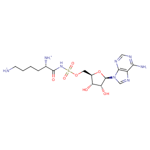
| HET Code: | KAA |
|---|---|
| Formula: | C16H27N8O7S |
| Molecular weight: | 475.500 g/mol |
| DrugBank ID: | - |
| Buried Surface Area: | 72.05 % |
| Polar Surface area: | 257.5 Å2 |
| Number of | |
|---|---|
| H-Bond Acceptors: | 11 |
| H-Bond Donors: | 5 |
| Rings: | 3 |
| Aromatic rings: | 2 |
| Anionic atoms: | 1 |
| Cationic atoms: | 2 |
| Rule of Five Violation: | 1 |
| Rotatable Bonds: | 10 |
| X | Y | Z |
|---|---|---|
| 0.0208125 | 26.8415 | 31.5464 |
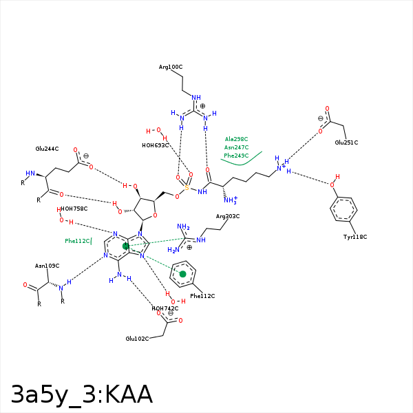


Represent the protein/ligand binding mode, centered on the ligand
Dashed lines represents hydrogen bonds and metal interactions
Green residue labels for amino acids with hydrophobic contacts (green lines) to the ligand
| Ligand | Protein | Interaction | |||
|---|---|---|---|---|---|
| Atom | Atom | Residue | Distance (Å) | Angle (°) | Type |
| NZ | OE2 | GLU- 78 | 3.17 | 122.04 | H-Bond (Ligand Donor) |
| NZ | OE1 | GLU- 78 | 3.43 | 131.59 | H-Bond (Ligand Donor) |
| NZ | OE2 | GLU- 78 | 3.17 | 0 | Ionic (Ligand Cationic) |
| NZ | OE1 | GLU- 78 | 3.43 | 0 | Ionic (Ligand Cationic) |
| N | OE1 | GLU- 78 | 3.49 | 0 | Ionic (Ligand Cationic) |
| O | NH2 | ARG- 100 | 2.79 | 157.87 | H-Bond (Protein Donor) |
| O1S | NH1 | ARG- 100 | 2.89 | 173.88 | H-Bond (Protein Donor) |
| N6 | OE1 | GLU- 102 | 2.9 | 147.3 | H-Bond (Ligand Donor) |
| N1 | N | ASN- 109 | 3.27 | 161.02 | H-Bond (Protein Donor) |
| N6 | O | ASN- 109 | 3.12 | 126.65 | H-Bond (Ligand Donor) |
| C1' | CE2 | PHE- 112 | 3.71 | 0 | Hydrophobic |
| DuAr | DuAr | PHE- 112 | 3.51 | 0 | Aromatic Face/Face |
| C5' | CE | MET- 114 | 3.7 | 0 | Hydrophobic |
| C4' | SD | MET- 114 | 3.76 | 0 | Hydrophobic |
| C1' | SD | MET- 114 | 4.4 | 0 | Hydrophobic |
| NZ | OH | TYR- 118 | 2.97 | 168.69 | H-Bond (Ligand Donor) |
| O3' | OE2 | GLU- 244 | 2.77 | 158.22 | H-Bond (Ligand Donor) |
| O2' | O | GLU- 244 | 3.18 | 175.25 | H-Bond (Ligand Donor) |
| CG | CZ | PHE- 249 | 3.48 | 0 | Hydrophobic |
| NZ | OE2 | GLU- 251 | 2.71 | 154.51 | H-Bond (Ligand Donor) |
| NZ | OE1 | GLU- 251 | 3.46 | 148.31 | H-Bond (Ligand Donor) |
| NZ | OE2 | GLU- 251 | 2.71 | 0 | Ionic (Ligand Cationic) |
| NZ | OE1 | GLU- 251 | 3.46 | 0 | Ionic (Ligand Cationic) |
| C5' | CB | ALA- 298 | 3.89 | 0 | Hydrophobic |
| CB | CB | ALA- 298 | 3.66 | 0 | Hydrophobic |
| O2' | N | GLY- 300 | 3.19 | 120.08 | H-Bond (Protein Donor) |
| C2' | CD | ARG- 303 | 4.12 | 0 | Hydrophobic |
| O2S | O | HOH- 693 | 2.66 | 123.85 | H-Bond (Protein Donor) |
| N7 | O | HOH- 742 | 3.01 | 179.96 | H-Bond (Protein Donor) |
| N3 | O | HOH- 758 | 2.93 | 165.22 | H-Bond (Protein Donor) |
