sc-PDB
An Annotated Database of Druggable Binding Sites from the Protein DataBank
An Annotated Database of Druggable Binding Sites from the Protein DataBank
1.800 Å
X-ray
2007-04-19
| Name: | Phosphoribosylamine--glycine ligase |
|---|---|
| ID: | PUR2_AQUAE |
| AC: | O66949 |
| Organism: | Aquifex aeolicus |
| Reign: | Bacteria |
| TaxID: | 224324 |
| EC Number: | / |
| Chain Name: | Percentage of Residues within binding site |
|---|---|
| B | 100 % |
| B-Factor: | 19.267 |
|---|---|
| Number of residues: | 47 |
| Including | |
| Standard Amino Acids: | 41 |
| Non Standard Amino Acids: | 0 |
| Water Molecules: | 6 |
| Cofactors: | |
| Metals: | |
| Ligandability | Volume (Å3) |
|---|---|
| 0.870 | 1468.125 |
| % Hydrophobic | % Polar |
|---|---|
| 33.56 | 66.44 |
| According to VolSite | |
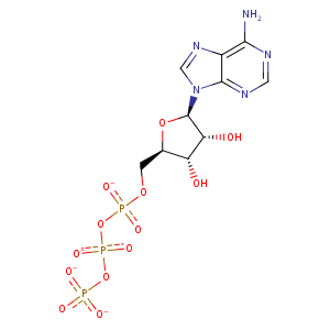
| HET Code: | ATP |
|---|---|
| Formula: | C10H12N5O13P3 |
| Molecular weight: | 503.149 g/mol |
| DrugBank ID: | DB00171 |
| Buried Surface Area: | 65.47 % |
| Polar Surface area: | 319.88 Å2 |
| Number of | |
|---|---|
| H-Bond Acceptors: | 17 |
| H-Bond Donors: | 3 |
| Rings: | 3 |
| Aromatic rings: | 2 |
| Anionic atoms: | 4 |
| Cationic atoms: | 0 |
| Rule of Five Violation: | 2 |
| Rotatable Bonds: | 8 |
| X | Y | Z |
|---|---|---|
| -28.0082 | 9.68123 | 44.5951 |
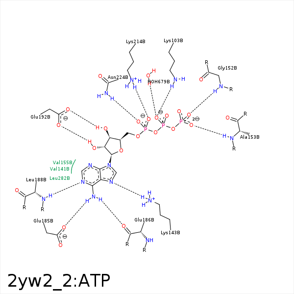


Represent the protein/ligand binding mode, centered on the ligand
Dashed lines represents hydrogen bonds and metal interactions
Green residue labels for amino acids with hydrophobic contacts (green lines) to the ligand
| Ligand | Protein | Interaction | |||
|---|---|---|---|---|---|
| Atom | Atom | Residue | Distance (Å) | Angle (°) | Type |
| O1B | NZ | LYS- 103 | 2.73 | 153.59 | H-Bond (Protein Donor) |
| N7 | NZ | LYS- 143 | 3.03 | 140.92 | H-Bond (Protein Donor) |
| O2G | N | GLY- 150 | 3.33 | 123.26 | H-Bond (Protein Donor) |
| O2G | N | GLY- 152 | 2.95 | 144.54 | H-Bond (Protein Donor) |
| O3G | N | ALA- 153 | 2.93 | 150.22 | H-Bond (Protein Donor) |
| C1' | CG2 | VAL- 155 | 4.12 | 0 | Hydrophobic |
| N6 | OE2 | GLU- 185 | 2.78 | 163.93 | H-Bond (Ligand Donor) |
| N6 | O | GLU- 186 | 3.06 | 137.42 | H-Bond (Ligand Donor) |
| N1 | N | LEU- 188 | 2.92 | 173.99 | H-Bond (Protein Donor) |
| O3' | OE1 | GLU- 192 | 2.64 | 169.39 | H-Bond (Ligand Donor) |
| O2' | OE1 | GLU- 192 | 3.28 | 135.11 | H-Bond (Ligand Donor) |
| O2' | OE2 | GLU- 192 | 2.51 | 161.1 | H-Bond (Ligand Donor) |
| O1A | NZ | LYS- 214 | 3.87 | 0 | Ionic (Protein Cationic) |
| O2A | NZ | LYS- 214 | 3.17 | 0 | Ionic (Protein Cationic) |
| O2A | NZ | LYS- 214 | 3.17 | 160.83 | H-Bond (Protein Donor) |
| O1A | ND2 | ASN- 224 | 3.24 | 145.66 | H-Bond (Protein Donor) |
| O5' | ND2 | ASN- 224 | 3.2 | 136.23 | H-Bond (Protein Donor) |
| C2' | CD1 | LEU- 282 | 3.73 | 0 | Hydrophobic |
| O1B | O | HOH- 679 | 2.92 | 163.13 | H-Bond (Protein Donor) |
