sc-PDB
An Annotated Database of Druggable Binding Sites from the Protein DataBank
An Annotated Database of Druggable Binding Sites from the Protein DataBank
2.010 Å
X-ray
2011-05-06
| Name: | Pteridine reductase |
|---|---|
| ID: | O76290_TRYBB |
| AC: | O76290 |
| Organism: | Trypanosoma brucei brucei |
| Reign: | Eukaryota |
| TaxID: | 5702 |
| EC Number: | / |
| Chain Name: | Percentage of Residues within binding site |
|---|---|
| B | 4 % |
| C | 96 % |
| B-Factor: | 23.214 |
|---|---|
| Number of residues: | 25 |
| Including | |
| Standard Amino Acids: | 22 |
| Non Standard Amino Acids: | 2 |
| Water Molecules: | 1 |
| Cofactors: | NDP |
| Metals: | |
| Ligandability | Volume (Å3) |
|---|---|
| 0.769 | 772.875 |
| % Hydrophobic | % Polar |
|---|---|
| 48.47 | 51.53 |
| According to VolSite | |
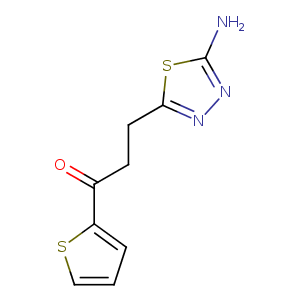
| HET Code: | WHF |
|---|---|
| Formula: | C9H9N3OS2 |
| Molecular weight: | 239.317 g/mol |
| DrugBank ID: | - |
| Buried Surface Area: | 74.3 % |
| Polar Surface area: | 125.35 Å2 |
| Number of | |
|---|---|
| H-Bond Acceptors: | 4 |
| H-Bond Donors: | 1 |
| Rings: | 2 |
| Aromatic rings: | 2 |
| Anionic atoms: | 0 |
| Cationic atoms: | 0 |
| Rule of Five Violation: | 0 |
| Rotatable Bonds: | 4 |
| X | Y | Z |
|---|---|---|
| 0.5446 | 15.1676 | 30.3917 |
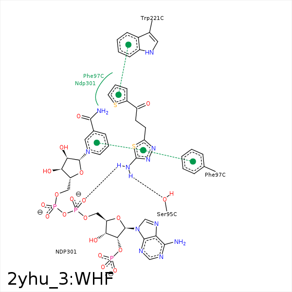


Represent the protein/ligand binding mode, centered on the ligand
Dashed lines represents hydrogen bonds and metal interactions
Green residue labels for amino acids with hydrophobic contacts (green lines) to the ligand
| Ligand | Protein | Interaction | |||
|---|---|---|---|---|---|
| Atom | Atom | Residue | Distance (Å) | Angle (°) | Type |
| NAA | OG | SER- 95 | 3.12 | 149.35 | H-Bond (Ligand Donor) |
| CAF | CZ | PHE- 97 | 4.35 | 0 | Hydrophobic |
| CAG | CE2 | PHE- 97 | 3.95 | 0 | Hydrophobic |
| SAK | CB | PHE- 97 | 4.01 | 0 | Hydrophobic |
| DuAr | DuAr | PHE- 97 | 3.84 | 0 | Aromatic Face/Face |
| SAJ | CG2 | VAL- 206 | 3.57 | 0 | Hydrophobic |
| CAG | CG | PRO- 210 | 4.18 | 0 | Hydrophobic |
| SAJ | CE2 | TRP- 221 | 3.43 | 0 | Hydrophobic |
| CAF | C4N | NDP- 301 | 3.69 | 0 | Hydrophobic |
| CAG | C3N | NDP- 301 | 4.1 | 0 | Hydrophobic |
| NAA | O2D | NDP- 301 | 3.37 | 123.55 | H-Bond (Ligand Donor) |
| NAA | O2A | NDP- 301 | 2.93 | 153.25 | H-Bond (Ligand Donor) |
