sc-PDB
An Annotated Database of Druggable Binding Sites from the Protein DataBank
An Annotated Database of Druggable Binding Sites from the Protein DataBank
2.150 Å
X-ray
2011-03-18
| Name: | 2',3'-cyclic-nucleotide 3'-phosphodiesterase |
|---|---|
| ID: | CN37_MOUSE |
| AC: | P16330 |
| Organism: | Mus musculus |
| Reign: | Eukaryota |
| TaxID: | 10090 |
| EC Number: | 3.1.4.37 |
| Chain Name: | Percentage of Residues within binding site |
|---|---|
| A | 100 % |
| B-Factor: | 31.834 |
|---|---|
| Number of residues: | 23 |
| Including | |
| Standard Amino Acids: | 19 |
| Non Standard Amino Acids: | 0 |
| Water Molecules: | 4 |
| Cofactors: | |
| Metals: | |
| Ligandability | Volume (Å3) |
|---|---|
| 0.196 | 526.500 |
| % Hydrophobic | % Polar |
|---|---|
| 43.59 | 56.41 |
| According to VolSite | |
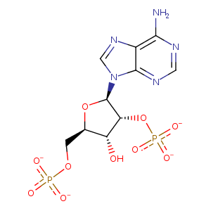
| HET Code: | NAP |
|---|---|
| Formula: | C21H25N7O17P3 |
| Molecular weight: | 740.381 g/mol |
| DrugBank ID: | DB03461 |
| Buried Surface Area: | 40.3 % |
| Polar Surface area: | 405.54 Å2 |
| Number of | |
|---|---|
| H-Bond Acceptors: | 21 |
| H-Bond Donors: | 5 |
| Rings: | 5 |
| Aromatic rings: | 3 |
| Anionic atoms: | 4 |
| Cationic atoms: | 1 |
| Rule of Five Violation: | 2 |
| Rotatable Bonds: | 13 |
| X | Y | Z |
|---|---|---|
| -7.2723 | 4.47204 | -25.1832 |
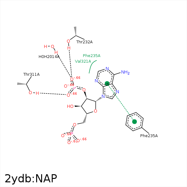


Represent the protein/ligand binding mode, centered on the ligand
Dashed lines represents hydrogen bonds and metal interactions
Green residue labels for amino acids with hydrophobic contacts (green lines) to the ligand
| Ligand | Protein | Interaction | |||
|---|---|---|---|---|---|
| Atom | Atom | Residue | Distance (Å) | Angle (°) | Type |
| C1B | CD2 | TYR- 168 | 4.17 | 0 | Hydrophobic |
| C4B | CE2 | TYR- 168 | 3.67 | 0 | Hydrophobic |
| O2B | NE2 | HIS- 230 | 3.37 | 155.64 | H-Bond (Protein Donor) |
| O3X | NE2 | HIS- 230 | 3.47 | 120.88 | H-Bond (Protein Donor) |
| O3X | OG1 | THR- 232 | 3.19 | 161.07 | H-Bond (Protein Donor) |
| C1B | CD1 | PHE- 235 | 4.14 | 0 | Hydrophobic |
| DuAr | DuAr | PHE- 235 | 3.72 | 0 | Aromatic Face/Face |
| O1X | OG1 | THR- 311 | 2.62 | 155.78 | H-Bond (Protein Donor) |
| C3B | CG | PRO- 320 | 3.69 | 0 | Hydrophobic |
| O3X | O | HOH- 2014 | 2.71 | 179.96 | H-Bond (Protein Donor) |
