sc-PDB
An Annotated Database of Druggable Binding Sites from the Protein DataBank
An Annotated Database of Druggable Binding Sites from the Protein DataBank
2.340 Å
X-ray
2010-10-13
| Name: | Guanine nucleotide-binding protein alpha-1 subunit |
|---|---|
| ID: | GPA1_ARATH |
| AC: | P18064 |
| Organism: | Arabidopsis thaliana |
| Reign: | Eukaryota |
| TaxID: | 3702 |
| EC Number: | / |
| Chain Name: | Percentage of Residues within binding site |
|---|---|
| C | 100 % |
| B-Factor: | 49.655 |
|---|---|
| Number of residues: | 44 |
| Including | |
| Standard Amino Acids: | 39 |
| Non Standard Amino Acids: | 2 |
| Water Molecules: | 3 |
| Cofactors: | |
| Metals: | MG |
| Ligandability | Volume (Å3) |
|---|---|
| 0.414 | 891.000 |
| % Hydrophobic | % Polar |
|---|---|
| 43.56 | 56.44 |
| According to VolSite | |
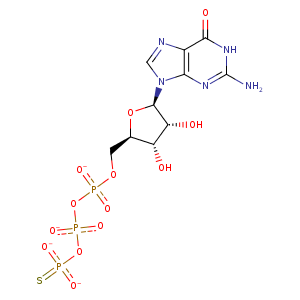
| HET Code: | GSP |
|---|---|
| Formula: | C10H14N5O13P3S |
| Molecular weight: | 537.230 g/mol |
| DrugBank ID: | DB01864 |
| Buried Surface Area: | 77.56 % |
| Polar Surface area: | 344.91 Å2 |
| Number of | |
|---|---|
| H-Bond Acceptors: | 17 |
| H-Bond Donors: | 6 |
| Rings: | 3 |
| Aromatic rings: | 1 |
| Anionic atoms: | 2 |
| Cationic atoms: | 0 |
| Rule of Five Violation: | 3 |
| Rotatable Bonds: | 8 |
| X | Y | Z |
|---|---|---|
| -26.1148 | -70.8562 | -11.9326 |


Represent the protein/ligand binding mode, centered on the ligand
Dashed lines represents hydrogen bonds and metal interactions
Green residue labels for amino acids with hydrophobic contacts (green lines) to the ligand
| Ligand | Protein | Interaction | |||
|---|---|---|---|---|---|
| Atom | Atom | Residue | Distance (Å) | Angle (°) | Type |
| O3B | N | GLU- 48 | 2.93 | 140.53 | H-Bond (Protein Donor) |
| C5' | CB | GLU- 48 | 4.17 | 0 | Hydrophobic |
| O1B | N | GLY- 50 | 3 | 146.94 | H-Bond (Protein Donor) |
| O3A | N | GLY- 50 | 3.31 | 139.01 | H-Bond (Protein Donor) |
| O2G | NZ | LYS- 51 | 2.63 | 170.89 | H-Bond (Protein Donor) |
| O1B | NZ | LYS- 51 | 2.78 | 141.01 | H-Bond (Protein Donor) |
| O1B | N | LYS- 51 | 2.91 | 148.51 | H-Bond (Protein Donor) |
| O2G | NZ | LYS- 51 | 2.63 | 0 | Ionic (Protein Cationic) |
| O1B | NZ | LYS- 51 | 2.78 | 0 | Ionic (Protein Cationic) |
| O2B | N | SER- 52 | 2.89 | 163 | H-Bond (Protein Donor) |
| O1A | N | THR- 53 | 2.93 | 140.01 | H-Bond (Protein Donor) |
| O1A | OG1 | THR- 53 | 2.61 | 164.35 | H-Bond (Protein Donor) |
| N2 | OD2 | ASP- 162 | 3.2 | 149.2 | H-Bond (Ligand Donor) |
| O2' | O | LEU- 187 | 2.66 | 155.88 | H-Bond (Ligand Donor) |
| O3' | O | TYR- 188 | 2.92 | 163.63 | H-Bond (Ligand Donor) |
| C3' | CB | ARG- 190 | 4.14 | 0 | Hydrophobic |
| C4' | CG | ARG- 190 | 4.31 | 0 | Hydrophobic |
| O3G | N | THR- 193 | 3.07 | 164.2 | H-Bond (Protein Donor) |
| O2G | N | GLY- 221 | 2.74 | 129.55 | H-Bond (Protein Donor) |
| N7 | ND2 | ASN- 287 | 3.05 | 142.07 | H-Bond (Protein Donor) |
| O4' | NZ | LYS- 288 | 3.19 | 138.48 | H-Bond (Protein Donor) |
| O6 | N | LYS- 288 | 3.24 | 126.89 | H-Bond (Protein Donor) |
| N1 | OD1 | ASP- 290 | 2.76 | 154.89 | H-Bond (Ligand Donor) |
| N2 | OD2 | ASP- 290 | 2.83 | 159.92 | H-Bond (Ligand Donor) |
| N2 | OD1 | ASP- 290 | 3.36 | 129.17 | H-Bond (Ligand Donor) |
| O6 | N | ALA- 355 | 2.93 | 139.34 | H-Bond (Protein Donor) |
| O3G | MG | MG- 1378 | 2.19 | 0 | Metal Acceptor |
| O2B | MG | MG- 1378 | 1.97 | 0 | Metal Acceptor |
| O2A | O | HOH- 2005 | 2.74 | 149.01 | H-Bond (Protein Donor) |
