sc-PDB
An Annotated Database of Druggable Binding Sites from the Protein DataBank
An Annotated Database of Druggable Binding Sites from the Protein DataBank
1.750 Å
X-ray
2009-12-24
| Name: | Prostaglandin reductase 3 |
|---|---|
| ID: | ZADH2_HUMAN |
| AC: | Q8N4Q0 |
| Organism: | Homo sapiens |
| Reign: | Eukaryota |
| TaxID: | 9606 |
| EC Number: | 1 |
| Chain Name: | Percentage of Residues within binding site |
|---|---|
| B | 100 % |
| B-Factor: | 16.072 |
|---|---|
| Number of residues: | 20 |
| Including | |
| Standard Amino Acids: | 19 |
| Non Standard Amino Acids: | 1 |
| Water Molecules: | 0 |
| Cofactors: | NAP |
| Metals: | |
| Ligandability | Volume (Å3) |
|---|---|
| 0.932 | 567.000 |
| % Hydrophobic | % Polar |
|---|---|
| 52.38 | 47.62 |
| According to VolSite | |
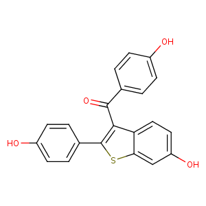
| HET Code: | X1H |
|---|---|
| Formula: | C22H16O4S |
| Molecular weight: | 376.425 g/mol |
| DrugBank ID: | - |
| Buried Surface Area: | 55.04 % |
| Polar Surface area: | 95 Å2 |
| Number of | |
|---|---|
| H-Bond Acceptors: | 4 |
| H-Bond Donors: | 2 |
| Rings: | 4 |
| Aromatic rings: | 4 |
| Anionic atoms: | 0 |
| Cationic atoms: | 0 |
| Rule of Five Violation: | 1 |
| Rotatable Bonds: | 4 |
| X | Y | Z |
|---|---|---|
| -37.6646 | -10.3883 | -46.7982 |
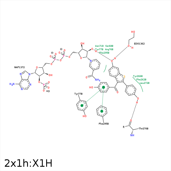


Represent the protein/ligand binding mode, centered on the ligand
Dashed lines represents hydrogen bonds and metal interactions
Green residue labels for amino acids with hydrophobic contacts (green lines) to the ligand
| Ligand | Protein | Interaction | |||
|---|---|---|---|---|---|
| Atom | Atom | Residue | Distance (Å) | Angle (°) | Type |
| O4 | OG | SER- 68 | 3.44 | 123.49 | H-Bond (Protein Donor) |
| C14 | CB | SER- 68 | 3.49 | 0 | Hydrophobic |
| C5 | CD | ARG- 76 | 4.18 | 0 | Hydrophobic |
| C13 | CZ | TYR- 77 | 3.43 | 0 | Hydrophobic |
| S1 | CZ | PHE- 262 | 3.82 | 0 | Hydrophobic |
| S1 | CE1 | TYR- 266 | 3.7 | 0 | Hydrophobic |
| O2 | O | THR- 270 | 3.15 | 151.79 | H-Bond (Ligand Donor) |
| C5 | CD2 | LEU- 272 | 3.67 | 0 | Hydrophobic |
| C20 | CZ | PHE- 295 | 3.4 | 0 | Hydrophobic |
| O4 | O2D | NAP- 1372 | 2.94 | 158.58 | H-Bond (Protein Donor) |
