sc-PDB
An Annotated Database of Druggable Binding Sites from the Protein DataBank
An Annotated Database of Druggable Binding Sites from the Protein DataBank
2.950 Å
X-ray
2008-08-08
| Name: | AcsD |
|---|---|
| ID: | Q93AT8_DICCH |
| AC: | Q93AT8 |
| Organism: | Dickeya chrysanthemi |
| Reign: | Bacteria |
| TaxID: | 556 |
| EC Number: | / |
| Chain Name: | Percentage of Residues within binding site |
|---|---|
| B | 100 % |
| B-Factor: | 18.222 |
|---|---|
| Number of residues: | 27 |
| Including | |
| Standard Amino Acids: | 27 |
| Non Standard Amino Acids: | 0 |
| Water Molecules: | 0 |
| Cofactors: | |
| Metals: | |
| Ligandability | Volume (Å3) |
|---|---|
| 0.541 | 1886.625 |
| % Hydrophobic | % Polar |
|---|---|
| 39.00 | 61.00 |
| According to VolSite | |
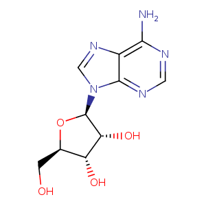
| HET Code: | ADN |
|---|---|
| Formula: | C10H13N5O4 |
| Molecular weight: | 267.241 g/mol |
| DrugBank ID: | DB00640 |
| Buried Surface Area: | 59.75 % |
| Polar Surface area: | 139.54 Å2 |
| Number of | |
|---|---|
| H-Bond Acceptors: | 8 |
| H-Bond Donors: | 4 |
| Rings: | 3 |
| Aromatic rings: | 2 |
| Anionic atoms: | 0 |
| Cationic atoms: | 0 |
| Rule of Five Violation: | 0 |
| Rotatable Bonds: | 2 |
| X | Y | Z |
|---|---|---|
| -8.91911 | 17.6597 | -8.89853 |
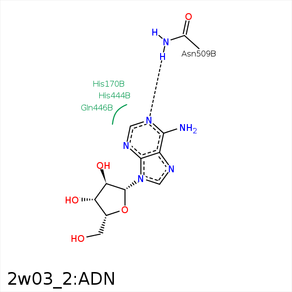


Represent the protein/ligand binding mode, centered on the ligand
Dashed lines represents hydrogen bonds and metal interactions
Green residue labels for amino acids with hydrophobic contacts (green lines) to the ligand
| Ligand | Protein | Interaction | |||
|---|---|---|---|---|---|
| Atom | Atom | Residue | Distance (Å) | Angle (°) | Type |
| DuAr | DuAr | HIS- 170 | 3.93 | 0 | Aromatic Face/Face |
| C4' | CB | SER- 279 | 3.87 | 0 | Hydrophobic |
| C2' | CG2 | THR- 301 | 3.69 | 0 | Hydrophobic |
| C1' | CG | GLN- 446 | 3.93 | 0 | Hydrophobic |
| C4' | CG | GLN- 446 | 3.81 | 0 | Hydrophobic |
| N1 | ND2 | ASN- 509 | 3.07 | 154.72 | H-Bond (Protein Donor) |
