sc-PDB
An Annotated Database of Druggable Binding Sites from the Protein DataBank
An Annotated Database of Druggable Binding Sites from the Protein DataBank
2.150 Å
X-ray
2007-11-22
| Name: | NTPase P4 |
|---|---|
| ID: | Q94M05_9VIRU |
| AC: | Q94M05 |
| Organism: | Pseudomonas phage phi12 |
| Reign: | Viruses |
| TaxID: | 161736 |
| EC Number: | / |
| Chain Name: | Percentage of Residues within binding site |
|---|---|
| B | 40 % |
| C | 60 % |
| B-Factor: | 51.133 |
|---|---|
| Number of residues: | 40 |
| Including | |
| Standard Amino Acids: | 34 |
| Non Standard Amino Acids: | 1 |
| Water Molecules: | 5 |
| Cofactors: | |
| Metals: | MG |
| Ligandability | Volume (Å3) |
|---|---|
| 0.466 | 702.000 |
| % Hydrophobic | % Polar |
|---|---|
| 37.50 | 62.50 |
| According to VolSite | |
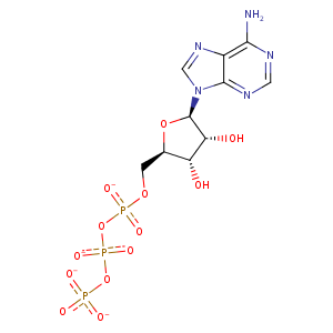
| HET Code: | ATP |
|---|---|
| Formula: | C10H12N5O13P3 |
| Molecular weight: | 503.149 g/mol |
| DrugBank ID: | DB00171 |
| Buried Surface Area: | 57.91 % |
| Polar Surface area: | 319.88 Å2 |
| Number of | |
|---|---|
| H-Bond Acceptors: | 17 |
| H-Bond Donors: | 3 |
| Rings: | 3 |
| Aromatic rings: | 2 |
| Anionic atoms: | 4 |
| Cationic atoms: | 0 |
| Rule of Five Violation: | 2 |
| Rotatable Bonds: | 8 |
| X | Y | Z |
|---|---|---|
| 32.3805 | 52.1936 | 27.2349 |
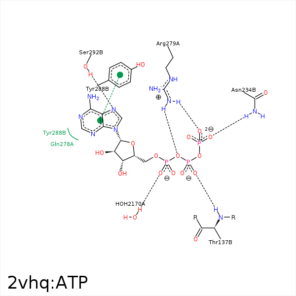


Represent the protein/ligand binding mode, centered on the ligand
Dashed lines represents hydrogen bonds and metal interactions
Green residue labels for amino acids with hydrophobic contacts (green lines) to the ligand
| Ligand | Protein | Interaction | |||
|---|---|---|---|---|---|
| Atom | Atom | Residue | Distance (Å) | Angle (°) | Type |
| O2B | N | THR- 137 | 2.92 | 161.59 | H-Bond (Protein Donor) |
| C5' | CG | PRO- 138 | 4.02 | 0 | Hydrophobic |
| O3G | ND2 | ASN- 234 | 2.75 | 140.66 | H-Bond (Protein Donor) |
| O1G | CZ | ARG- 272 | 3.81 | 0 | Ionic (Protein Cationic) |
| O2G | CZ | ARG- 279 | 3.65 | 0 | Ionic (Protein Cationic) |
| O1A | CZ | ARG- 279 | 3.75 | 0 | Ionic (Protein Cationic) |
| O1A | NH2 | ARG- 279 | 3.28 | 137.63 | H-Bond (Protein Donor) |
| O1A | NE | ARG- 279 | 3.41 | 135.35 | H-Bond (Protein Donor) |
| O3A | NH2 | ARG- 279 | 3.26 | 158.42 | H-Bond (Protein Donor) |
| C3' | CG | ARG- 279 | 4.48 | 0 | Hydrophobic |
| C1' | CZ | TYR- 288 | 4.1 | 0 | Hydrophobic |
| DuAr | DuAr | TYR- 288 | 3.48 | 0 | Aromatic Face/Face |
| N7 | OG | SER- 292 | 2.78 | 146.96 | H-Bond (Protein Donor) |
| O1G | MG | MG- 1328 | 2.32 | 0 | Metal Acceptor |
| O2B | MG | MG- 1328 | 2.4 | 0 | Metal Acceptor |
| O3B | MG | MG- 1328 | 2.79 | 0 | Metal Acceptor |
| O2A | MG | MG- 1328 | 2.03 | 0 | Metal Acceptor |
| O3G | O | HOH- 2129 | 3.44 | 179.98 | H-Bond (Protein Donor) |
| O1A | O | HOH- 2170 | 2.77 | 149.51 | H-Bond (Protein Donor) |
