sc-PDB
An Annotated Database of Druggable Binding Sites from the Protein DataBank
An Annotated Database of Druggable Binding Sites from the Protein DataBank
1.950 Å
X-ray
2007-09-27
| Name: | Hematopoietic prostaglandin D synthase |
|---|---|
| ID: | HPGDS_HUMAN |
| AC: | O60760 |
| Organism: | Homo sapiens |
| Reign: | Eukaryota |
| TaxID: | 9606 |
| EC Number: | / |
| Chain Name: | Percentage of Residues within binding site |
|---|---|
| B | 100 % |
| B-Factor: | 16.506 |
|---|---|
| Number of residues: | 17 |
| Including | |
| Standard Amino Acids: | 17 |
| Non Standard Amino Acids: | 0 |
| Water Molecules: | 0 |
| Cofactors: | |
| Metals: | |
| Ligandability | Volume (Å3) |
|---|---|
| 0.966 | 1539.000 |
| % Hydrophobic | % Polar |
|---|---|
| 39.25 | 60.75 |
| According to VolSite | |
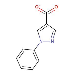
| HET Code: | ZZA |
|---|---|
| Formula: | C10H7N2O2 |
| Molecular weight: | 187.175 g/mol |
| DrugBank ID: | DB08790 |
| Buried Surface Area: | 59.32 % |
| Polar Surface area: | 57.95 Å2 |
| Number of | |
|---|---|
| H-Bond Acceptors: | 3 |
| H-Bond Donors: | 0 |
| Rings: | 2 |
| Aromatic rings: | 2 |
| Anionic atoms: | 1 |
| Cationic atoms: | 0 |
| Rule of Five Violation: | 0 |
| Rotatable Bonds: | 2 |
| X | Y | Z |
|---|---|---|
| 4.40329 | 15.1917 | 1.45679 |
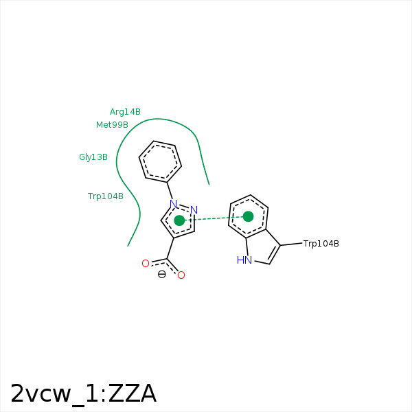


Represent the protein/ligand binding mode, centered on the ligand
Dashed lines represents hydrogen bonds and metal interactions
Green residue labels for amino acids with hydrophobic contacts (green lines) to the ligand
| Ligand | Protein | Interaction | |||
|---|---|---|---|---|---|
| Atom | Atom | Residue | Distance (Å) | Angle (°) | Type |
| C10 | CG | ARG- 14 | 3.53 | 0 | Hydrophobic |
| C11 | CB | MET- 99 | 3.45 | 0 | Hydrophobic |
| C13 | CE | MET- 99 | 4.16 | 0 | Hydrophobic |
| DuAr | DuAr | TRP- 104 | 3.69 | 0 | Aromatic Face/Face |
| C12 | SG | CYS- 156 | 4.11 | 0 | Hydrophobic |
