sc-PDB
An Annotated Database of Druggable Binding Sites from the Protein DataBank
An Annotated Database of Druggable Binding Sites from the Protein DataBank
1.900 Å
X-ray
2008-10-02
| Name: | TetR family transcriptional regulator |
|---|---|
| ID: | Q58L87_MYCSM |
| AC: | Q58L87 |
| Organism: | Mycobacterium smegmatis |
| Reign: | Bacteria |
| TaxID: | 1772 |
| EC Number: | / |
| Chain Name: | Percentage of Residues within binding site |
|---|---|
| A | 95 % |
| B | 5 % |
| B-Factor: | 22.942 |
|---|---|
| Number of residues: | 21 |
| Including | |
| Standard Amino Acids: | 21 |
| Non Standard Amino Acids: | 0 |
| Water Molecules: | 0 |
| Cofactors: | |
| Metals: | |
| Ligandability | Volume (Å3) |
|---|---|
| 0.877 | 1066.500 |
| % Hydrophobic | % Polar |
|---|---|
| 41.46 | 58.54 |
| According to VolSite | |
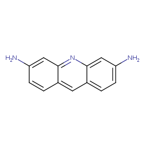
| HET Code: | PRL |
|---|---|
| Formula: | C13H11N3 |
| Molecular weight: | 209.247 g/mol |
| DrugBank ID: | DB01123 |
| Buried Surface Area: | 58.22 % |
| Polar Surface area: | 64.92 Å2 |
| Number of | |
|---|---|
| H-Bond Acceptors: | 3 |
| H-Bond Donors: | 2 |
| Rings: | 3 |
| Aromatic rings: | 3 |
| Anionic atoms: | 0 |
| Cationic atoms: | 0 |
| Rule of Five Violation: | 0 |
| Rotatable Bonds: | 0 |
| X | Y | Z |
|---|---|---|
| 7.28006 | -5.27075 | 6.67625 |
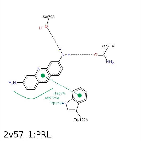


Represent the protein/ligand binding mode, centered on the ligand
Dashed lines represents hydrogen bonds and metal interactions
Green residue labels for amino acids with hydrophobic contacts (green lines) to the ligand
| Ligand | Protein | Interaction | |||
|---|---|---|---|---|---|
| Atom | Atom | Residue | Distance (Å) | Angle (°) | Type |
| N15 | OG | SER- 70 | 2.84 | 163.59 | H-Bond (Ligand Donor) |
| N15 | OD1 | ASN- 71 | 2.99 | 165.84 | H-Bond (Ligand Donor) |
| C3 | CD1 | ILE- 74 | 4.15 | 0 | Hydrophobic |
| C1 | CB | ASP- 125 | 3.52 | 0 | Hydrophobic |
| C5 | CB | TRP- 152 | 3.46 | 0 | Hydrophobic |
