sc-PDB
An Annotated Database of Druggable Binding Sites from the Protein DataBank
An Annotated Database of Druggable Binding Sites from the Protein DataBank
1.800 Å
X-ray
1997-03-08
| Name: | UDP-glucose 4-epimerase |
|---|---|
| ID: | GALE_ECOLI |
| AC: | P09147 |
| Organism: | Escherichia coli |
| Reign: | Bacteria |
| TaxID: | 83333 |
| EC Number: | 5.1.3.2 |
| Chain Name: | Percentage of Residues within binding site |
|---|---|
| A | 100 % |
| B-Factor: | 28.449 |
|---|---|
| Number of residues: | 35 |
| Including | |
| Standard Amino Acids: | 33 |
| Non Standard Amino Acids: | 1 |
| Water Molecules: | 1 |
| Cofactors: | NAD |
| Metals: | |
| Ligandability | Volume (Å3) |
|---|---|
| 1.118 | 1154.250 |
| % Hydrophobic | % Polar |
|---|---|
| 47.08 | 52.92 |
| According to VolSite | |
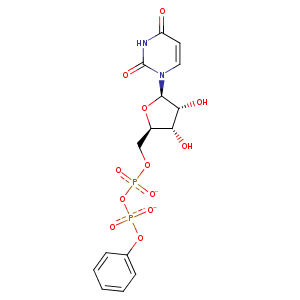
| HET Code: | UPP |
|---|---|
| Formula: | C15H16N2O12P2 |
| Molecular weight: | 478.241 g/mol |
| DrugBank ID: | DB02790 |
| Buried Surface Area: | 67.98 % |
| Polar Surface area: | 226.67 Å2 |
| Number of | |
|---|---|
| H-Bond Acceptors: | 12 |
| H-Bond Donors: | 3 |
| Rings: | 3 |
| Aromatic rings: | 1 |
| Anionic atoms: | 2 |
| Cationic atoms: | 0 |
| Rule of Five Violation: | 1 |
| Rotatable Bonds: | 8 |
| X | Y | Z |
|---|---|---|
| 18.3211 | 57.1298 | 9.41052 |
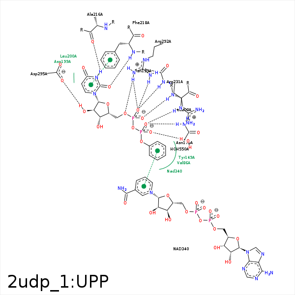


Represent the protein/ligand binding mode, centered on the ligand
Dashed lines represents hydrogen bonds and metal interactions
Green residue labels for amino acids with hydrophobic contacts (green lines) to the ligand
| Ligand | Protein | Interaction | |||
|---|---|---|---|---|---|
| Atom | Atom | Residue | Distance (Å) | Angle (°) | Type |
| C5' | CG2 | VAL- 86 | 3.79 | 0 | Hydrophobic |
| O1B | ND2 | ASN- 179 | 2.92 | 157.13 | H-Bond (Protein Donor) |
| O1A | ND2 | ASN- 199 | 3.13 | 149.61 | H-Bond (Protein Donor) |
| C1B | CD1 | LEU- 200 | 4.47 | 0 | Hydrophobic |
| C4B | CD2 | LEU- 200 | 4.41 | 0 | Hydrophobic |
| C5B | CB | LEU- 200 | 4.16 | 0 | Hydrophobic |
| O2A | N | LEU- 200 | 2.73 | 160.7 | H-Bond (Protein Donor) |
| N3 | O | ALA- 216 | 2.95 | 147.93 | H-Bond (Ligand Donor) |
| O2 | N | PHE- 218 | 3.03 | 163.58 | H-Bond (Protein Donor) |
| O1B | NE | ARG- 231 | 2.99 | 138.79 | H-Bond (Protein Donor) |
| O1B | CZ | ARG- 231 | 4 | 0 | Ionic (Protein Cationic) |
| C5B | CG | ARG- 231 | 4.03 | 0 | Hydrophobic |
| C1B | CG2 | VAL- 269 | 3.65 | 0 | Hydrophobic |
| C4B | CG2 | VAL- 269 | 4.14 | 0 | Hydrophobic |
| O5' | NH2 | ARG- 292 | 3.07 | 134.92 | H-Bond (Protein Donor) |
| O1A | NH2 | ARG- 292 | 2.84 | 150.41 | H-Bond (Protein Donor) |
| O1A | NH1 | ARG- 292 | 3.08 | 138.18 | H-Bond (Protein Donor) |
| O1A | CZ | ARG- 292 | 3.38 | 0 | Ionic (Protein Cationic) |
| O2B | O | HOH- 550 | 2.98 | 169.17 | H-Bond (Protein Donor) |
