sc-PDB
An Annotated Database of Druggable Binding Sites from the Protein DataBank
An Annotated Database of Druggable Binding Sites from the Protein DataBank
2.500 Å
X-ray
1997-01-23
| Name: | Tyrosine phenol-lyase |
|---|---|
| ID: | TPL_CITFR |
| AC: | P31013 |
| Organism: | Citrobacter freundii |
| Reign: | Bacteria |
| TaxID: | 546 |
| EC Number: | 4.1.99.2 |
| Chain Name: | Percentage of Residues within binding site |
|---|---|
| A | 18 % |
| B | 82 % |
| B-Factor: | 21.364 |
|---|---|
| Number of residues: | 22 |
| Including | |
| Standard Amino Acids: | 21 |
| Non Standard Amino Acids: | 1 |
| Water Molecules: | 0 |
| Cofactors: | |
| Metals: | |
| Ligandability | Volume (Å3) |
|---|---|
| 0.153 | 607.500 |
| % Hydrophobic | % Polar |
|---|---|
| 47.78 | 52.22 |
| According to VolSite | |
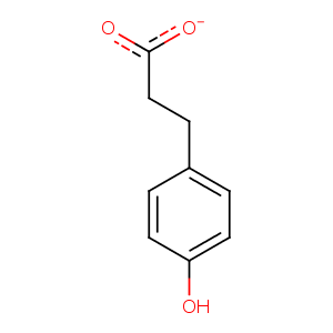
| HET Code: | HPP |
|---|---|
| Formula: | C9H9O3 |
| Molecular weight: | 165.166 g/mol |
| DrugBank ID: | DB03897 |
| Buried Surface Area: | 50.84 % |
| Polar Surface area: | 60.36 Å2 |
| Number of | |
|---|---|
| H-Bond Acceptors: | 3 |
| H-Bond Donors: | 1 |
| Rings: | 1 |
| Aromatic rings: | 1 |
| Anionic atoms: | 1 |
| Cationic atoms: | 0 |
| Rule of Five Violation: | 0 |
| Rotatable Bonds: | 3 |
| X | Y | Z |
|---|---|---|
| -23.2634 | 17.5466 | 3.92408 |
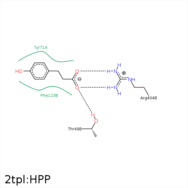


Represent the protein/ligand binding mode, centered on the ligand
Dashed lines represents hydrogen bonds and metal interactions
Green residue labels for amino acids with hydrophobic contacts (green lines) to the ligand
| Ligand | Protein | Interaction | |||
|---|---|---|---|---|---|
| Atom | Atom | Residue | Distance (Å) | Angle (°) | Type |
| O2 | OG1 | THR- 49 | 2.5 | 150.09 | H-Bond (Protein Donor) |
| C8 | CB | SER- 51 | 4.29 | 0 | Hydrophobic |
| C7 | CZ | TYR- 71 | 3.37 | 0 | Hydrophobic |
| C8 | CZ | PHE- 123 | 4.28 | 0 | Hydrophobic |
| C2 | CE | MET- 379 | 4.41 | 0 | Hydrophobic |
| O1 | NH2 | ARG- 404 | 3.34 | 139.23 | H-Bond (Protein Donor) |
| O1 | NH1 | ARG- 404 | 3.03 | 157.47 | H-Bond (Protein Donor) |
| O2 | NH2 | ARG- 404 | 3 | 147.37 | H-Bond (Protein Donor) |
| O1 | CZ | ARG- 404 | 3.61 | 0 | Ionic (Protein Cationic) |
| O2 | CZ | ARG- 404 | 3.9 | 0 | Ionic (Protein Cationic) |
