sc-PDB
An Annotated Database of Druggable Binding Sites from the Protein DataBank
An Annotated Database of Druggable Binding Sites from the Protein DataBank
1.900 Å
X-ray
2007-06-12
| Name: | Methionine aminopeptidase |
|---|---|
| ID: | MAP1_ECOLI |
| AC: | P0AE18 |
| Organism: | Escherichia coli |
| Reign: | Bacteria |
| TaxID: | 83333 |
| EC Number: | / |
| Chain Name: | Percentage of Residues within binding site |
|---|---|
| A | 100 % |
| B-Factor: | 18.138 |
|---|---|
| Number of residues: | 30 |
| Including | |
| Standard Amino Acids: | 28 |
| Non Standard Amino Acids: | 2 |
| Water Molecules: | 0 |
| Cofactors: | |
| Metals: | MN MN |
| Ligandability | Volume (Å3) |
|---|---|
| 0.609 | 479.250 |
| % Hydrophobic | % Polar |
|---|---|
| 40.14 | 59.86 |
| According to VolSite | |
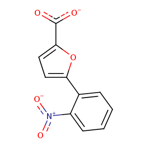
| HET Code: | B23 |
|---|---|
| Formula: | C11H6NO5 |
| Molecular weight: | 232.169 g/mol |
| DrugBank ID: | DB07408 |
| Buried Surface Area: | 71.94 % |
| Polar Surface area: | 99.09 Å2 |
| Number of | |
|---|---|
| H-Bond Acceptors: | 4 |
| H-Bond Donors: | 0 |
| Rings: | 2 |
| Aromatic rings: | 2 |
| Anionic atoms: | 2 |
| Cationic atoms: | 1 |
| Rule of Five Violation: | 0 |
| Rotatable Bonds: | 3 |
| X | Y | Z |
|---|---|---|
| -3.15806 | -0.0728235 | 9.08253 |
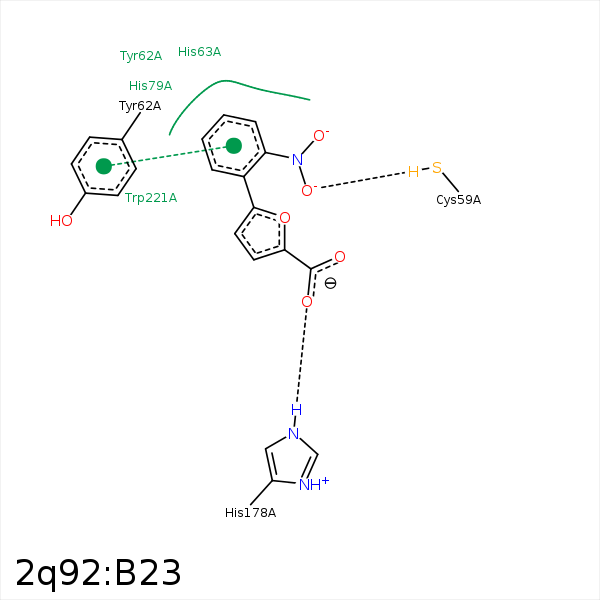


Represent the protein/ligand binding mode, centered on the ligand
Dashed lines represents hydrogen bonds and metal interactions
Green residue labels for amino acids with hydrophobic contacts (green lines) to the ligand
| Ligand | Protein | Interaction | |||
|---|---|---|---|---|---|
| Atom | Atom | Residue | Distance (Å) | Angle (°) | Type |
| CAK | CB | TYR- 62 | 4.45 | 0 | Hydrophobic |
| CAI | CD2 | TYR- 62 | 3.48 | 0 | Hydrophobic |
| CAI | CB | HIS- 63 | 3.81 | 0 | Hydrophobic |
| CAK | CB | TYR- 65 | 4.34 | 0 | Hydrophobic |
| OAO | NE2 | HIS- 178 | 2.8 | 141.22 | H-Bond (Protein Donor) |
| OAM | NE2 | HIS- 178 | 3.23 | 125.92 | H-Bond (Protein Donor) |
| CAK | CZ3 | TRP- 221 | 3.39 | 0 | Hydrophobic |
| OAN | MN | MN- 300 | 2.33 | 0 | Metal Acceptor |
| OAO | MN | MN- 300 | 2.23 | 0 | Metal Acceptor |
| OAN | MN | MN- 301 | 2.15 | 0 | Metal Acceptor |
