sc-PDB
An Annotated Database of Druggable Binding Sites from the Protein DataBank
An Annotated Database of Druggable Binding Sites from the Protein DataBank
2.100 Å
X-ray
2007-04-20
| Name: | 5-methylthioadenosine/S-adenosylhomocysteine deaminase |
|---|---|
| ID: | MTAD_THEMA |
| AC: | Q9X034 |
| Organism: | Thermotoga maritima |
| Reign: | Bacteria |
| TaxID: | 243274 |
| EC Number: | 3.5.4.28 |
| Chain Name: | Percentage of Residues within binding site |
|---|---|
| A | 100 % |
| B-Factor: | 29.051 |
|---|---|
| Number of residues: | 35 |
| Including | |
| Standard Amino Acids: | 31 |
| Non Standard Amino Acids: | 1 |
| Water Molecules: | 3 |
| Cofactors: | |
| Metals: | ZN |
| Ligandability | Volume (Å3) |
|---|---|
| 0.455 | 526.500 |
| % Hydrophobic | % Polar |
|---|---|
| 55.77 | 44.23 |
| According to VolSite | |
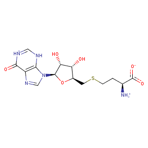
| HET Code: | SIB |
|---|---|
| Formula: | C14H19N5O6S |
| Molecular weight: | 385.396 g/mol |
| DrugBank ID: | - |
| Buried Surface Area: | 75.54 % |
| Polar Surface area: | 202.03 Å2 |
| Number of | |
|---|---|
| H-Bond Acceptors: | 9 |
| H-Bond Donors: | 4 |
| Rings: | 3 |
| Aromatic rings: | 1 |
| Anionic atoms: | 1 |
| Cationic atoms: | 1 |
| Rule of Five Violation: | 1 |
| Rotatable Bonds: | 7 |
| X | Y | Z |
|---|---|---|
| 45.4167 | 31.9868 | -1.48492 |
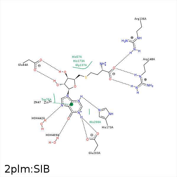


Represent the protein/ligand binding mode, centered on the ligand
Dashed lines represents hydrogen bonds and metal interactions
Green residue labels for amino acids with hydrophobic contacts (green lines) to the ligand
| Ligand | Protein | Interaction | |||
|---|---|---|---|---|---|
| Atom | Atom | Residue | Distance (Å) | Angle (°) | Type |
| C2' | SD | MET- 60 | 3.96 | 0 | Hydrophobic |
| C1' | CD1 | LEU- 76 | 4.47 | 0 | Hydrophobic |
| O3' | OE1 | GLU- 84 | 2.71 | 168.54 | H-Bond (Ligand Donor) |
| O2' | OE1 | GLU- 84 | 3.28 | 120.53 | H-Bond (Ligand Donor) |
| O2' | OE2 | GLU- 84 | 2.72 | 174.71 | H-Bond (Ligand Donor) |
| C5' | SD | MET- 114 | 3.91 | 0 | Hydrophobic |
| C5' | CD1 | TYR- 115 | 4.28 | 0 | Hydrophobic |
| C3' | CE1 | TYR- 115 | 3.88 | 0 | Hydrophobic |
| O | CZ | ARG- 136 | 3.69 | 0 | Ionic (Protein Cationic) |
| O | NH1 | ARG- 136 | 3.04 | 144.83 | H-Bond (Protein Donor) |
| CB | CG1 | VAL- 139 | 4.32 | 0 | Hydrophobic |
| OXT | CZ | ARG- 148 | 3.8 | 0 | Ionic (Protein Cationic) |
| O | CZ | ARG- 148 | 3.51 | 0 | Ionic (Protein Cationic) |
| OXT | NH2 | ARG- 148 | 3.03 | 156.07 | H-Bond (Protein Donor) |
| O | NE | ARG- 148 | 2.75 | 171.18 | H-Bond (Protein Donor) |
| O | NH2 | ARG- 148 | 3.41 | 129.92 | H-Bond (Protein Donor) |
| N3 | NE2 | HIS- 173 | 3.03 | 149.25 | H-Bond (Ligand Donor) |
| O6 | O | HOH- 409 | 2.94 | 121.69 | H-Bond (Protein Donor) |
| N7 | O | HOH- 442 | 2.72 | 179.96 | H-Bond (Protein Donor) |
