sc-PDB
An Annotated Database of Druggable Binding Sites from the Protein DataBank
An Annotated Database of Druggable Binding Sites from the Protein DataBank
1.600 Å
X-ray
2007-02-09
| Name: | Pyridoxamine 5'-phosphate oxidase-related FMN-binding protein |
|---|---|
| ID: | Q28VU1_JANSC |
| AC: | Q28VU1 |
| Organism: | Jannaschia sp. |
| Reign: | Bacteria |
| TaxID: | 290400 |
| EC Number: | / |
| Chain Name: | Percentage of Residues within binding site |
|---|---|
| A | 70 % |
| B | 30 % |
| B-Factor: | 18.750 |
|---|---|
| Number of residues: | 36 |
| Including | |
| Standard Amino Acids: | 33 |
| Non Standard Amino Acids: | 0 |
| Water Molecules: | 3 |
| Cofactors: | |
| Metals: | |
| Ligandability | Volume (Å3) |
|---|---|
| 1.111 | 1177.875 |
| % Hydrophobic | % Polar |
|---|---|
| 38.40 | 61.60 |
| According to VolSite | |
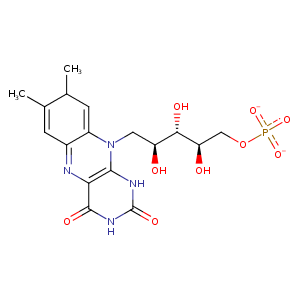
| HET Code: | FMN |
|---|---|
| Formula: | C17H19N4O9P |
| Molecular weight: | 454.328 g/mol |
| DrugBank ID: | DB03247 |
| Buried Surface Area: | 66.04 % |
| Polar Surface area: | 217.05 Å2 |
| Number of | |
|---|---|
| H-Bond Acceptors: | 12 |
| H-Bond Donors: | 4 |
| Rings: | 3 |
| Aromatic rings: | 1 |
| Anionic atoms: | 2 |
| Cationic atoms: | 0 |
| Rule of Five Violation: | 1 |
| Rotatable Bonds: | 7 |
| X | Y | Z |
|---|---|---|
| 33.1369 | 39.7361 | 80.1988 |
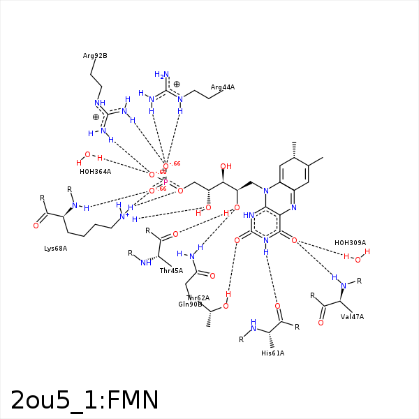


Represent the protein/ligand binding mode, centered on the ligand
Dashed lines represents hydrogen bonds and metal interactions
Green residue labels for amino acids with hydrophobic contacts (green lines) to the ligand
| Ligand | Protein | Interaction | |||
|---|---|---|---|---|---|
| Atom | Atom | Residue | Distance (Å) | Angle (°) | Type |
| O3P | CZ | ARG- 44 | 3.28 | 0 | Ionic (Protein Cationic) |
| O3P | NE | ARG- 44 | 3.02 | 138.41 | H-Bond (Protein Donor) |
| O3P | NH2 | ARG- 44 | 2.69 | 152.92 | H-Bond (Protein Donor) |
| C2' | CG | ARG- 44 | 4.02 | 0 | Hydrophobic |
| C7M | CG2 | THR- 45 | 4.03 | 0 | Hydrophobic |
| C8 | CG2 | THR- 45 | 3.96 | 0 | Hydrophobic |
| C8M | CB | THR- 45 | 3.83 | 0 | Hydrophobic |
| O2' | O | THR- 45 | 2.75 | 175 | H-Bond (Ligand Donor) |
| O4 | N | VAL- 47 | 3.03 | 172.59 | H-Bond (Protein Donor) |
| N3 | O | HIS- 61 | 3.12 | 166.4 | H-Bond (Ligand Donor) |
| O2 | OG1 | THR- 62 | 2.61 | 160.57 | H-Bond (Protein Donor) |
| O2 | NZ | LYS- 68 | 3.25 | 139.45 | H-Bond (Protein Donor) |
| O4' | NZ | LYS- 68 | 2.62 | 124.55 | H-Bond (Protein Donor) |
| O5' | NZ | LYS- 68 | 3.14 | 137.14 | H-Bond (Protein Donor) |
| O1P | N | LYS- 68 | 2.85 | 150.23 | H-Bond (Protein Donor) |
| O1P | NZ | LYS- 68 | 2.75 | 145.32 | H-Bond (Protein Donor) |
| O1P | NZ | LYS- 68 | 2.75 | 0 | Ionic (Protein Cationic) |
| C7M | CZ2 | TRP- 83 | 3.75 | 0 | Hydrophobic |
| C8M | CE3 | TRP- 83 | 3.76 | 0 | Hydrophobic |
| C8M | CB | GLN- 90 | 3.77 | 0 | Hydrophobic |
| O2' | NE2 | GLN- 90 | 3.02 | 160.6 | H-Bond (Protein Donor) |
| O5' | NH1 | ARG- 92 | 3.31 | 126.75 | H-Bond (Protein Donor) |
| O2P | NH1 | ARG- 92 | 3.33 | 170.05 | H-Bond (Protein Donor) |
| O3P | NH2 | ARG- 92 | 2.99 | 146.17 | H-Bond (Protein Donor) |
| O3P | CZ | ARG- 92 | 3.82 | 0 | Ionic (Protein Cationic) |
| C1' | CD2 | LEU- 151 | 4.03 | 0 | Hydrophobic |
| C8 | CD2 | LEU- 151 | 4.31 | 0 | Hydrophobic |
| C7M | CD2 | LEU- 153 | 4.09 | 0 | Hydrophobic |
| C8 | CD2 | LEU- 153 | 3.8 | 0 | Hydrophobic |
| O4 | O | HOH- 309 | 2.74 | 179.97 | H-Bond (Protein Donor) |
| O2P | O | HOH- 364 | 2.56 | 179.95 | H-Bond (Protein Donor) |
