sc-PDB
An Annotated Database of Druggable Binding Sites from the Protein DataBank
An Annotated Database of Druggable Binding Sites from the Protein DataBank
1.800 Å
X-ray
1993-08-24
| Name: | Alcohol dehydrogenase E chain |
|---|---|
| ID: | ADH1E_HORSE |
| AC: | P00327 |
| Organism: | Equus caballus |
| Reign: | Eukaryota |
| TaxID: | 9796 |
| EC Number: | 1.1.1.1 |
| Chain Name: | Percentage of Residues within binding site |
|---|---|
| A | 98 % |
| B | 2 % |
| B-Factor: | 19.521 |
|---|---|
| Number of residues: | 58 |
| Including | |
| Standard Amino Acids: | 52 |
| Non Standard Amino Acids: | 1 |
| Water Molecules: | 5 |
| Cofactors: | |
| Metals: | ZN |
| Ligandability | Volume (Å3) |
|---|---|
| 1.097 | 931.500 |
| % Hydrophobic | % Polar |
|---|---|
| 50.72 | 49.28 |
| According to VolSite | |
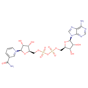
| HET Code: | NAD |
|---|---|
| Formula: | C21H26N7O14P2 |
| Molecular weight: | 662.417 g/mol |
| DrugBank ID: | - |
| Buried Surface Area: | 70.71 % |
| Polar Surface area: | 343.54 Å2 |
| Number of | |
|---|---|
| H-Bond Acceptors: | 18 |
| H-Bond Donors: | 6 |
| Rings: | 5 |
| Aromatic rings: | 3 |
| Anionic atoms: | 2 |
| Cationic atoms: | 1 |
| Rule of Five Violation: | 3 |
| Rotatable Bonds: | 11 |
| X | Y | Z |
|---|---|---|
| 0.122114 | 15.5138 | 15.1958 |


Represent the protein/ligand binding mode, centered on the ligand
Dashed lines represents hydrogen bonds and metal interactions
Green residue labels for amino acids with hydrophobic contacts (green lines) to the ligand
| Ligand | Protein | Interaction | |||
|---|---|---|---|---|---|
| Atom | Atom | Residue | Distance (Å) | Angle (°) | Type |
| C5N | SG | CYS- 46 | 3.94 | 0 | Hydrophobic |
| O2A | NH2 | ARG- 47 | 2.9 | 140.34 | H-Bond (Protein Donor) |
| O2A | NE | ARG- 47 | 2.79 | 149.87 | H-Bond (Protein Donor) |
| O1N | N | ARG- 47 | 3.13 | 160.48 | H-Bond (Protein Donor) |
| O2A | CZ | ARG- 47 | 3.27 | 0 | Ionic (Protein Cationic) |
| C3D | CG | ARG- 47 | 4.01 | 0 | Hydrophobic |
| C2D | CB | ARG- 47 | 4.05 | 0 | Hydrophobic |
| O2D | OG | SER- 48 | 2.81 | 159.91 | H-Bond (Ligand Donor) |
| O3D | NE2 | HIS- 51 | 3.19 | 167.85 | H-Bond (Protein Donor) |
| C5N | SG | CYS- 174 | 3.33 | 0 | Hydrophobic |
| C4N | CG2 | THR- 178 | 3.54 | 0 | Hydrophobic |
| O2N | N | VAL- 203 | 3 | 169.36 | H-Bond (Protein Donor) |
| C5D | CB | VAL- 203 | 4.43 | 0 | Hydrophobic |
| C5N | CG2 | VAL- 203 | 3.88 | 0 | Hydrophobic |
| O3B | OD2 | ASP- 223 | 2.68 | 140.62 | H-Bond (Ligand Donor) |
| O2B | OD1 | ASP- 223 | 2.74 | 168.26 | H-Bond (Ligand Donor) |
| O3B | NZ | LYS- 228 | 2.74 | 143.46 | H-Bond (Protein Donor) |
| C5D | CG1 | VAL- 268 | 4.21 | 0 | Hydrophobic |
| C1B | CG1 | ILE- 269 | 4.4 | 0 | Hydrophobic |
| O3D | O | ILE- 269 | 2.64 | 165.41 | H-Bond (Ligand Donor) |
| C3N | CG1 | VAL- 292 | 4.43 | 0 | Hydrophobic |
| N7N | O | VAL- 292 | 2.97 | 173.7 | H-Bond (Ligand Donor) |
| O3D | N | VAL- 294 | 3.19 | 147.01 | H-Bond (Protein Donor) |
| C2D | CG2 | VAL- 294 | 4.43 | 0 | Hydrophobic |
| N7N | O | ALA- 317 | 3.09 | 157.14 | H-Bond (Ligand Donor) |
| O7N | N | PHE- 319 | 2.87 | 166.57 | H-Bond (Protein Donor) |
| O1N | NH1 | ARG- 369 | 2.88 | 147.51 | H-Bond (Protein Donor) |
| O1N | CZ | ARG- 369 | 3.9 | 0 | Ionic (Protein Cationic) |
| O1A | O | HOH- 436 | 2.77 | 136.39 | H-Bond (Protein Donor) |
| O1A | O | HOH- 476 | 2.73 | 162.45 | H-Bond (Protein Donor) |
| O2N | O | HOH- 477 | 2.73 | 179.97 | H-Bond (Protein Donor) |
