sc-PDB
An Annotated Database of Druggable Binding Sites from the Protein DataBank
An Annotated Database of Druggable Binding Sites from the Protein DataBank
2.530 Å
X-ray
2006-12-20
| Name: | 6,7-dimethyl-8-ribityllumazine synthase 2 |
|---|---|
| ID: | RISB2_RHILO |
| AC: | Q986N2 |
| Organism: | Rhizobium loti |
| Reign: | Bacteria |
| TaxID: | 266835 |
| EC Number: | 2.5.1.78 |
| Chain Name: | Percentage of Residues within binding site |
|---|---|
| H | 32 % |
| I | 68 % |
| B-Factor: | 21.766 |
|---|---|
| Number of residues: | 34 |
| Including | |
| Standard Amino Acids: | 34 |
| Non Standard Amino Acids: | 0 |
| Water Molecules: | 0 |
| Cofactors: | |
| Metals: | |
| Ligandability | Volume (Å3) |
|---|---|
| 1.212 | 1076.625 |
| % Hydrophobic | % Polar |
|---|---|
| 46.39 | 53.61 |
| According to VolSite | |
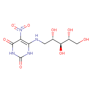
| HET Code: | INI |
|---|---|
| Formula: | C9H14N4O8 |
| Molecular weight: | 306.229 g/mol |
| DrugBank ID: | DB04162 |
| Buried Surface Area: | 63.49 % |
| Polar Surface area: | 196.96 Å2 |
| Number of | |
|---|---|
| H-Bond Acceptors: | 9 |
| H-Bond Donors: | 7 |
| Rings: | 1 |
| Aromatic rings: | 0 |
| Anionic atoms: | 1 |
| Cationic atoms: | 1 |
| Rule of Five Violation: | 2 |
| Rotatable Bonds: | 7 |
| X | Y | Z |
|---|---|---|
| 45.5687 | -11.9404 | 9.1461 |
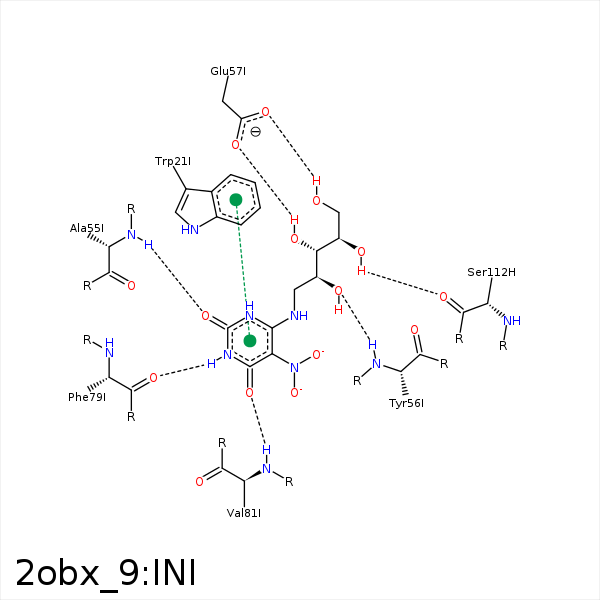


Represent the protein/ligand binding mode, centered on the ligand
Dashed lines represents hydrogen bonds and metal interactions
Green residue labels for amino acids with hydrophobic contacts (green lines) to the ligand
| Ligand | Protein | Interaction | |||
|---|---|---|---|---|---|
| Atom | Atom | Residue | Distance (Å) | Angle (°) | Type |
| O2 | N | ALA- 55 | 2.85 | 142.44 | H-Bond (Protein Donor) |
| O9 | N | TYR- 56 | 2.91 | 158.48 | H-Bond (Protein Donor) |
| C12 | CD2 | TYR- 56 | 4.38 | 0 | Hydrophobic |
| C9 | CB | TYR- 56 | 3.35 | 0 | Hydrophobic |
| O10 | OE1 | GLU- 57 | 3.02 | 147.39 | H-Bond (Ligand Donor) |
| N3 | O | PHE- 79 | 2.89 | 162.33 | H-Bond (Ligand Donor) |
| O4 | N | VAL- 81 | 2.82 | 140.74 | H-Bond (Protein Donor) |
| C8 | CG1 | VAL- 91 | 4.47 | 0 | Hydrophobic |
| C12 | CB | LEU- 111 | 3.76 | 0 | Hydrophobic |
| O11 | O | SER- 112 | 3 | 159.92 | H-Bond (Ligand Donor) |
| C12 | CB | SER- 112 | 3.97 | 0 | Hydrophobic |
| O12 | N | SER- 112 | 3.39 | 144.32 | H-Bond (Protein Donor) |
