sc-PDB
An Annotated Database of Druggable Binding Sites from the Protein DataBank
An Annotated Database of Druggable Binding Sites from the Protein DataBank
2.600 Å
X-ray
2005-12-22
| Name: | Alpha-hemolysin translocation ATP-binding protein HlyB |
|---|---|
| ID: | HLYBP_ECOLX |
| AC: | P08716 |
| Organism: | Escherichia coli |
| Reign: | Bacteria |
| TaxID: | 562 |
| EC Number: | / |
| Chain Name: | Percentage of Residues within binding site |
|---|---|
| C | 61 % |
| D | 39 % |
| B-Factor: | 12.973 |
|---|---|
| Number of residues: | 36 |
| Including | |
| Standard Amino Acids: | 36 |
| Non Standard Amino Acids: | 0 |
| Water Molecules: | 0 |
| Cofactors: | |
| Metals: | |
| Ligandability | Volume (Å3) |
|---|---|
| 0.574 | 1927.125 |
| % Hydrophobic | % Polar |
|---|---|
| 42.56 | 57.44 |
| According to VolSite | |
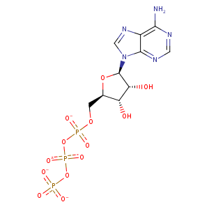
| HET Code: | ATP |
|---|---|
| Formula: | C10H12N5O13P3 |
| Molecular weight: | 503.149 g/mol |
| DrugBank ID: | DB00171 |
| Buried Surface Area: | 60.49 % |
| Polar Surface area: | 319.88 Å2 |
| Number of | |
|---|---|
| H-Bond Acceptors: | 17 |
| H-Bond Donors: | 3 |
| Rings: | 3 |
| Aromatic rings: | 2 |
| Anionic atoms: | 4 |
| Cationic atoms: | 0 |
| Rule of Five Violation: | 2 |
| Rotatable Bonds: | 8 |
| X | Y | Z |
|---|---|---|
| 21.8446 | 43.8585 | 17.8821 |
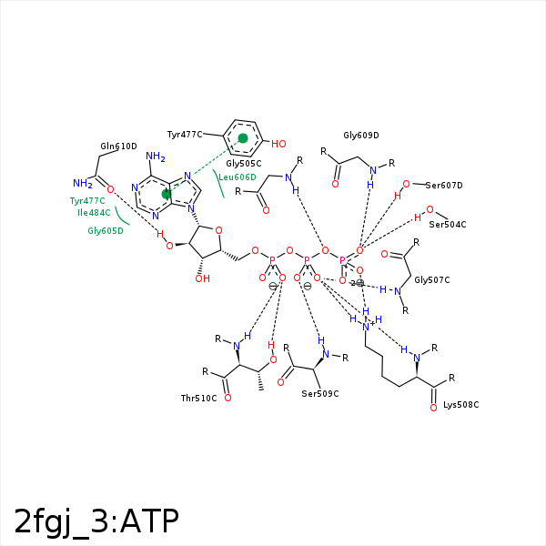


Represent the protein/ligand binding mode, centered on the ligand
Dashed lines represents hydrogen bonds and metal interactions
Green residue labels for amino acids with hydrophobic contacts (green lines) to the ligand
| Ligand | Protein | Interaction | |||
|---|---|---|---|---|---|
| Atom | Atom | Residue | Distance (Å) | Angle (°) | Type |
| C1' | CE1 | TYR- 477 | 4.24 | 0 | Hydrophobic |
| DuAr | DuAr | TYR- 477 | 3.53 | 0 | Aromatic Face/Face |
| C1' | CD1 | ILE- 484 | 3.87 | 0 | Hydrophobic |
| C4' | CG2 | ILE- 484 | 3.9 | 0 | Hydrophobic |
| O1G | OG | SER- 504 | 2.96 | 173.04 | H-Bond (Protein Donor) |
| O3B | N | GLY- 505 | 2.75 | 154.6 | H-Bond (Protein Donor) |
| O1B | N | SER- 506 | 3.17 | 126.57 | H-Bond (Protein Donor) |
| O1B | N | GLY- 507 | 3.11 | 153.28 | H-Bond (Protein Donor) |
| O3A | N | GLY- 507 | 3.29 | 132.52 | H-Bond (Protein Donor) |
| O2G | NZ | LYS- 508 | 2.82 | 159.68 | H-Bond (Protein Donor) |
| O1B | NZ | LYS- 508 | 2.6 | 140.78 | H-Bond (Protein Donor) |
| O1B | N | LYS- 508 | 3.16 | 152.05 | H-Bond (Protein Donor) |
| O2G | NZ | LYS- 508 | 2.82 | 0 | Ionic (Protein Cationic) |
| O1B | NZ | LYS- 508 | 2.6 | 0 | Ionic (Protein Cationic) |
| O2B | N | SER- 509 | 3.11 | 160.37 | H-Bond (Protein Donor) |
| O2A | OG1 | THR- 510 | 2.55 | 154.25 | H-Bond (Protein Donor) |
| O2A | N | THR- 510 | 2.91 | 164.17 | H-Bond (Protein Donor) |
| N1 | N | GLY- 605 | 3.22 | 136.31 | H-Bond (Protein Donor) |
| O1G | OG | SER- 607 | 3.12 | 148.51 | H-Bond (Protein Donor) |
| C2' | CB | SER- 607 | 4.43 | 0 | Hydrophobic |
| O1G | N | GLY- 609 | 2.78 | 129.78 | H-Bond (Protein Donor) |
| O2' | OE1 | GLN- 610 | 2.73 | 160.63 | H-Bond (Ligand Donor) |
